Orthopaedic implants have been tremendously successful in dealing with a host of skeletal diseases and trauma. The most important orthopaedic devices in use today are total joint replacements, which are used primarily in the replacement of hip and knee joints. Combined, hip and knee replacement surgery exceeds one million cases per year in the US: about 50% of the world market.
These medical devices have provided tremendous relief from pain for millions of patients suffering from osteoarthritis, rheumatoid arthritis, avascular necrosis, trauma and several other conditions.
They are long lived, typically expected to last ten years or more, and restore a level of function that many of these patients have not had for years. However, there are still problems.
Biomaterial requirements
Orthopaedic implants are currently made from synthetic materials typically consisting of alloys of cobalt, stainless steel and/or titanium, and polymers primarily consisting of ultra-high molecular-weight polyethylene, (UHMWPE) and poly(methyl methacrylate), otherwise known as acrylic bone cement (or PMMA).
In some instances, ceramic materials, typically alumina, are used in high-wearing locations. These materials are man-made and are meant to be permanently implanted in the body for the life of the patient.
How well do you really know your competitors?
Access the most comprehensive Company Profiles on the market, powered by GlobalData. Save hours of research. Gain competitive edge.

Thank you!
Your download email will arrive shortly
Not ready to buy yet? Download a free sample
We are confident about the unique quality of our Company Profiles. However, we want you to make the most beneficial decision for your business, so we offer a free sample that you can download by submitting the below form
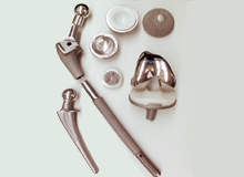
By GlobalDataThe typical hip implant consists of a femoral component and the acetabular component. Current joint replacement design has the femoral component made in a modular design with the ball separate from the stem of the overall component. The ball is typically made from highly wear-resistant biomaterials like cobalt-chromium-molybdenum alloy (Co-Cr-Mo). The stem can be made typically from titanium-aluminum-vanadium (Ti-6Al-4V), or other titanium alloy, Co-Cr-Mo, or stainless steel. Occasionally, the head may consist of a ceramic: alumina (Al2O3), or zirconia (ZrO2).
The femoral component is implanted inside the femur either by direct contact of the metal component with bone (biological fixation), or by use of bone cement. Biological fixation can take weeks or months to be fully complete, whereas bone cement sets in ten to 20 minutes and flows into the contours of the bone. In both cases the bone and the biomaterial form a close interface with a small amount of fibrous scar tissue between them. This interface is essential for the long-term success of the device.
The acetabular component typically consists of UHMWPE which articulates with the head of the femoral component to make up the joint. This cup is captured and held in place by a metal backing component that is usually attached to the bone by biological fixation. Total knee replacement implants similarly have two sides – a metallic femoral component (typically made from Co-Cr-Mo) and a tibial component (UHMWPE articulating surface) attached to a metallic component (tibial tray) that is, itself, typically cemented to the tibia by bone cement. Examples of these joint replacements can be seen in Figure 1.
These joint replacements experience significant loading and are exposed to wear-like processes, corrosion and environmental attack, and a host body that is trying its best to break up or expel the implant or its degradation products. The environment that orthopaedic implants experience is an important consideration and can be separated into three types: mechanical, bioelectrochemical and biological.
Mechanical environment
A typical person standing on one foot places about three times their body weight on the head of a total hip replacement. With more vigorous activities the load levels increase to well above three times body weight. These loads get transferred through the implant and then from the implant to the skeletal system. This mechanical environment can become very complex and adversely affect the implant, the adjacent bone, the bone cement or other elements of the system.
When high numbers of cyclic loads are applied, as is the case with most orthopaedic devices, fatigue failure becomes a critical concern.
A fatigue failure results when a component fractures by the accumulated effects of millions of cycles of loading, which damages the material to the point that a crack initiates and then propagates to failure over many cycles. Fatigue is the primary cause of implant fracture since patients typically load their implants up to several million cycles per year. An insidious aspect of fatigue failures is that the implant will appear to work well right up to the final applied load and then will fail without warning.
The mechanical environment can lead to a variety of problems that are generally categorised as implant loosening. The major reason why implants get revised is due to aseptic loosening. If the implant is cemented in place, then the loosening is often associated with fatigue failure of the bone cement, as well as stress shielding bone resorption. If the implant is biologically fixed, then loosening has to do with high levels of motion between the implant and the bone (so-called micromotion) and the development of a fibrous capsule that allows the implant to move relative to the bone.
Another important element of the mechanical environment is that there are cyclic wear processes that take place at many interfaces of the device. Wear occurs primarily at the articulating surfaces. The consequence of wear is the generation of micron and smaller-scale particles which are shed from the wearing surfaces of these biomaterials and accumulate in the surrounding tissue. Wear can also occur between metal and polymer interfaces, bone and metal, bone and bone cement, and at modular metal-metal connections.
These wear particles, whether they are UHMWPE, metal, ceramic or bone cement, are known to elicit a severe adverse biological reaction. The body’s immune and inflammatory systems respond by attempting to eliminate the particles. In bony sites, this process is autocatalytic and can lead to a process known as osteolysis.
Osteolysis (literally, bone destruction) is thought to be primarily a response to the presence of UHMWPE particles. However, osteolysis has been seen in the absence of UHMWPE in response to metal particles, bone cement and other particles. The consequence of osteolysis is that a lesion forms in the bone where there is a loss of strength and softening of the bone. If left unchecked, this site will eventually suffer a fracture and loss of implant stability.
One relatively recent advance in particle reduction has come with the development of cross-linked UHMWPE. When high-energy radiation interacts with UHMWPE in the absence of oxygen, a change in the polymer structure occurs. Cross-linking causes the long polymer chains to form bonds. Cross-linking reduces many of the important mechanical properties of the polymer, but also dramatically raises the wear resistance, almost eliminating the generation of particles. For some applications like hip replacement, cross-linked UHMWPE has significantly reduced particle generation and it is expected to lower particle-induced osteolysis accordingly.
Corrosion and body interaction
The composition of the human body, essentially ‘a bag of salt water’, can lead to corrosion of metals and degradation of polymers and ceramics. There are also proteins, enzymes, cytokines and a whole host of other biologically derived molecules that interact with and may degrade the materials, causing corrosion, oxidation and degradation.
Orthopaedic implant alloys are some of the most corrosion-resistant alloys known to modern engineering. They have survived in patients for many decades with little to no evidence of corrosion attack. This does not mean, however, that corrosion cannot happen with these alloys. The factors that increase corrosion rates usually include some form of mechanical abrasion of the oxide, a local environment that is removed from the normal body environment and the possibility of local accumulation of cell-derived biochemical species (inflammatory chemicals like hydrogen peroxide, hypochlorous acid) that when present in combination can lead to corrosion.
It is important to note that any combination of currently used orthopaedic alloys, when combined into a modular taper, is susceptible to severe corrosion attack. Even when Ti-6Al-4V modular taper interfaces are present, severe pitting and generalised corrosion of these alloys can occur, as shown in Figure 2.
There are biological consequences to increased corrosion that are not well appreciated or understood. Mechanically assisted corrosion can act to alter the voltage of the surface of a metal. With increased corrosion rates the voltage of the implant alloy becomes more negative. It is also the case that most of the biological processes associated with living cells are redox (corrosion-like) reactions. When an implant is corroding, it is possible that the voltage changes at the implant surface may affect the local cell behaviour and cause adverse effects.
For the past several decades the primary concern has been the release of metal ions that are thought to be toxic. Clearly the generation and movement of these ions are important and can have adverse biological effects both local to the implant and more systemically. However, some of these alternative effects are only now beginning to be appreciated.
Biological effects: infection
One of the most difficult aspects of orthopaedic implantation is the development of implant-centred infection. When this occurs, bacterial colonies form on the implant surface and they are impossible to eliminate without implant removal. Infection rates in joint replacements have remained roughly the same for the past several decades with little improvement seen.
The current practice for infected total joints is to surgically remove the implant from the site, to surgically debride the site to remove any retained bacterial colonies, and then to place a temporary implant made from bone cement and antibiotic which is left in the site for several weeks to months to kill any residual infection. Once the infection is resolved, a new total joint replacement is implanted. This process, while reasonably successful in eliminating the infection, is lengthy, painful and leaves the joint compromised.
Future directions
Total joint replacements are highly successful and restore pain-free mobility to millions of patients every year. While there are continuing problems, the overall message is that joint replacement surgery is a great success. However, there is still room for improvement.
Advances in materials technology that will address some of the limitations of current materials are expected. Improved wear surfaces, better understanding and control of corrosion and fatigue are all possible with the appropriate innovation of biomaterials and design.
Also, the future may bring advances in ‘smart prostheses’ where the device has elements in it that can sense, communicate and act upon the local body environment. Implants that can respond to adverse effects are also possible – releasing antibiotics if an infection is detected, for example.
There is much improvement possible to make implants that last more than the assumed ten years told to most patients today to more like 30–40 years. Much has been learned about how total joint replacements perform, but there is much yet to do in terms of innovation in materials, designs and approaches to orthopaedic devices.