Unlike a traditional 2D printer, which prints on a flat surface, 3D printers add another dimension – depth. Known as additive manufacturing, 3D printings distributes different materials up and down, left and right, back and forward, to print an item later by layer.
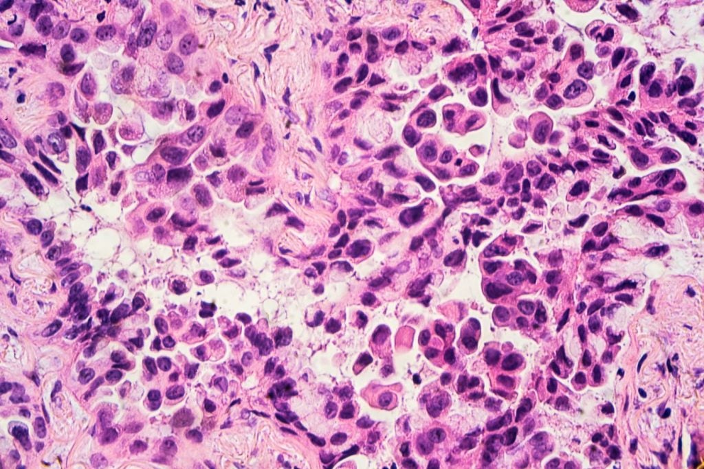
Bioprinting refers to 3D printers which deposit layers of biomaterial to build complex bodily structures like skin, bones and even corneas. The requisite cells are taken from a patient – or, if this isn’t possible, adult stem cells can be used – and cultivated into a bioink to ‘print’ an organic object. These are typically held together through some sort of dissolvable gel or collagen scaffold which can support the cells and mould them into the correct shape.
Go deeper with GlobalData

Printing body parts may well be the next step in organ transplantation – harvesting stem cells from a transplant recipient and printing them into a replacement organ could help bypass complications associated with organ transplant such as long waits for a suitable donor or immune rejection of the new organ.
Here is a look at some of the biggest breakthroughs in 3D bioprinting, and the various techniques used to create different body parts.
Bones: a regenerative structure for long-term healing
A team from Swansea University in the UK has developed a bioprinting process which can create an artificial bone matrix, using durable, regenerative biomaterial.
Currently, extremely complex bone fractures are treated through a surgical procedure called bone grafting, which replaces missing or damaged bones with synthetic, cement-based materials. However, this technique comes with its limitations, as these structures can often have inappropriate mechanical integrity and don’t allow the formation of new bone tissues.
How well do you really know your competitors?
Access the most comprehensive Company Profiles on the market, powered by GlobalData. Save hours of research. Gain competitive edge.

Thank you!
Your download email will arrive shortly
Not ready to buy yet? Download a free sample
We are confident about the unique quality of our Company Profiles. However, we want you to make the most beneficial decision for your business, so we offer a free sample that you can download by submitting the below form
By GlobalDataThe bioprinted bones can be printed in the exact structure needed with a durable and regenerative biomaterial. This material is made from gelatine, agarose, collagen alginate, calcium phosphate and polycaprolactone.
The bioprinted bone material is capable of fusing with a patient’s natural bones over time, eventually being replaced by them.
Corneas: a boost for biocompatibility
3D printed artificial corneas have been developed by a group of researchers in South Korea. The prototype corneas have been printed from biocompatible decellularized corneal stroma and stem cells.
The team behind the corneas hopes to see its invention replace the use of donor and synthetic corneas in surgery for cataracts and other sight complications.
Artificial corneas currently on the market are made up of recombinant collagen or chemical substances like synthetic polymer, which means they can often resist full incorporation into the eye or are not transparent after transplant.
The 3D printed corneas are constructed to mimic the lattice pattern of collagen fibrils within natural corneas by using the shear stress generated by the frictional force of the 3D printing process. Regulating the shear stress allowed the researchers to control the pattern in which the fibrils were printed, meaning they could make sure these artificial corneas adequately reflected the structure of the native human cornea.
Cartilage: revolutionising joint care
Researchers at an Australian biofabrication centre called BioFAB3D have built a handheld cartilage printing device called the BioPen.
The BioPen is filled with stem cells derived from a patient’s fat, which can create and surgically implant custom scaffolds of living material into failing joints. Much like 3D printed bones, the cartilage undergoes a process of growth and development within the body. So far it’s only been tested on sheep, but its developers hope that in future the BioPen can help to accelerate the regeneration of functional cartilage in human patients.
In practice, a surgeon will be able to use the 3D printing technology of the handheld stylus to place the bioink into a damaged joint layer by layer. The ink layers, which are comprised of stem cells and specially-selected growth factors within a biopolymer matrix, form a structure which a patient’s reproducing cells will rapidly strengthen for lasting cartilage repair.
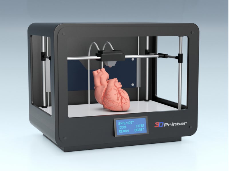
Hearts: beating the functionality challenge
A group of scientists at the American Friends of Tel Aviv University have 3D printed a fully-vascularised heart using fat tissue cells from a donor.
The fat cells were partially cultured and reprogrammed into heart cells. The entire heart structure is present with all cells, blood vessels, ventricles and chambers. The structure was based on medical images of the donor patient’s own heart.
The technology is still in its early stages – the heart the researchers have printed is the size of a rabbit’s heart and is unable to pump any blood. The researchers hope to test these printed hearts in animals once they have worked on rescaling the organs and getting them to beat.
Skin: a great leap in grafting for burn patients
Wake Forest School of Medicine has designed a printer that can print skin cells directly on to a burn wound.
The traditional treatment for severe burns is skin grafting, where healthy skin is harvested from an unburnt part of a patient’s body. This in itself can be traumatic to heal from, and in some cases there isn’t enough healthy skin left on the body to use.
In this printing technique, a patch of skin only 10% of the size of the burn can be used to grow enough cells for 3D printing. A scanner is used to determine the size and depth of the wound, and the printer then takes this information and prints hypodermic, dermic, and epidermic skin cells at the corresponding depths to cover the wound.
As trials around this technology move forward, the research team is hoping to see if stem cells from amniotic fluid and placentas are as effective as patient skin in healing these wounds.