A study has shown that interactive virtual reality (VR) can help physicians better prepare for complex surgeries and improve their confidence.
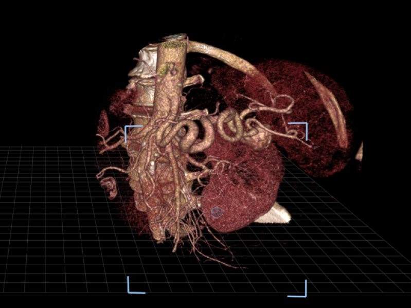
New VR technology can turn a patient’s pre-procedural CT scans into 3D images that the radiologist can virtually move and examine while wearing VR glasses.
Go deeper with GlobalData

Manipulating these images in a three-dimensional space allows physicians to familiarise themselves with a patient’s unique anatomical makeup.
Researchers from Stanford University School of Medicine compared this technology with a commonly-used visualisation software system that displays images on a standard two-dimensional platform.
In the study, three radiologists used both technologies to independently evaluate 17 splenic artery aneurysms in 14 patients. Researchers measured the radiologists’ accuracy in identifying inflow and outflow arteries associated with the aneurysms with each method.
The radiologists in the study also ranked improvements in their confidence on a four-point scale when using VR compared to the standard method.
How well do you really know your competitors?
Access the most comprehensive Company Profiles on the market, powered by GlobalData. Save hours of research. Gain competitive edge.

Thank you!
Your download email will arrive shortly
Not ready to buy yet? Download a free sample
We are confident about the unique quality of our Company Profiles. However, we want you to make the most beneficial decision for your business, so we offer a free sample that you can download by submitting the below form
By GlobalDataWhile accuracy was similar with both methods, confidence improved substantially with VR, with 93% indicating higher confidence in their abilities.
“Treating splenic artery aneurysms can be very difficult because of their intricate nature and anatomic variations from patient to patient,” said collaborating author on the study Zlatko Devcic.
“This new platform allows you to view a patient’s arterial anatomy in a three-dimensional image, as if it is right in front of you, which may help interventional radiologists more quickly and thoroughly plan for the equipment and tools they’ll need for a successful outcome.”
The researchers hope that future studies will explore the technology’s potential to reduce the time it takes to perform treatments.
“Pre-operative planning is possibly the most important step towards successfully treating a patient, so the value of VR cannot be understated,” said Devcic. “This technology gives us a totally different way to look at that structure and safely plan our approach to patient care.”
The research was presented at the Society of Interventional Radiology’s 2018 Annual Scientific Meeting.