
A team of researchers from Massachusetts Institute of Technology (MIT) and Massachusetts General Hospital (MGH) have created a new imaging system aid in the surgical removal of ovarian tumours.
Considered as one of the most difficult-to-treat tumours, ovarian cancer is commonly diagnosed in advanced stages when tumours have spread across the abdomen. Some tumours are too small to be identified by surgeons.
Go deeper with GlobalData

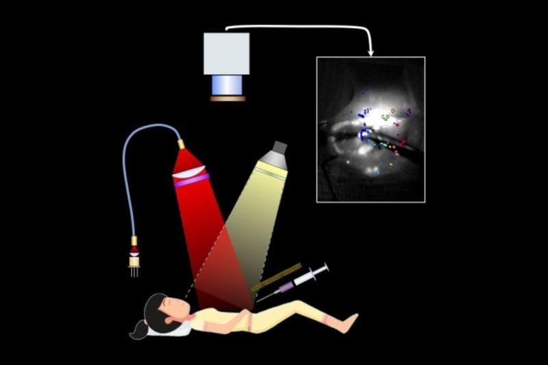
The new imaging system uses florescent light to visualise even the smallest tumours and offers continuous real-time images of the abdomen, paving way for improved accuracy of the surgery.
The system consists of chemical probes that release fluorescent light upon illumination with a laser. These probes are coated with a peptide that binds to the SPARC protein overexpressed by invasive ovarian cancer cells.
Tumours become florescent when the probes bind to them, making it easy for surgeons to spot them. When tested in mice, surgeons were able to find and remove tumours as small as 0.3mm.
According to the researchers, mice that underwent surgery guided by the new imaging system did not have any detectable tumours at ten days after surgery, while those in non-image-guided surgery group had multiple residual tumours.
How well do you really know your competitors?
Access the most comprehensive Company Profiles on the market, powered by GlobalData. Save hours of research. Gain competitive edge.

Thank you!
Your download email will arrive shortly
Not ready to buy yet? Download a free sample
We are confident about the unique quality of our Company Profiles. However, we want you to make the most beneficial decision for your business, so we offer a free sample that you can download by submitting the below form
By GlobalDataMGH medical gynaecologic oncology former director Michael Birrer said: “We know that the amount of tumour removed at the time of surgery for patients with advanced-stage ovarian cancer is directly correlated with their outcome.
“This imaging device will now allow the surgeon to go beyond the limits of resecting tumours visible to the naked eye, and should usher in a new age of effective debulking surgery.”
Mice that underwent the image-guided surgery had 40% longer median survival rate. However, the tumours had grown back by three weeks after the surgery.
As most patients require chemotherapy after surgery, the researchers intend to test the same approach in a separate preclinical study.
The researchers also plan to seek US regulatory approval for a Phase I clinical trial to assess the imaging system in human patients.
Moreover, they hope that the system could help in monitoring patients at risk of cancer recurrence in the future and eventually be used as a diagnostic for early detection of ovarian cancer.
