
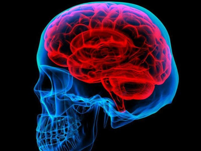
A team of researchers at the University of Southern California (USC) Viterbi School of Engineering has created a real-time thermal imaging method that could help doctors deliver fast, safe and precise thermal ablation treatments for numerous ailments including tumours and epilepsy.
Am emerging technique to destroy brain tumours uses a needle thin probe that can deliver radio frequency waves directly to a tumour, burning the tissue at temperatures of up to 60 degrees Celsius until it is destroyed. This technique is known as radiofrequency ablation (RFA) and is a minimally-invasive procedure.
Go deeper with GlobalData

USC research assistant professor John Stang said: “Although ablation is becoming increasingly popular, there is still no thermal imaging technology in regular clinical use to monitor these procedures in real time and ensure that the correct thermal dose is delivered the first time.”
Stang worked alongside professor and director of the USC Microwave Systems, Sensors, and Imaging Lab Mahta Moghaddam to develop the real time thermal imaging method and device that could fill this gap in the thermal imaging market. They published their findings in IEEE Transactions on Biomedical Engineering,
Moghaddam said: “Without real-time monitoring, there is the potential for both under-treatment and over-treatment. If there is under-treatment, doctors must perform additional rounds of thermal ablation until all of the tumour is destroyed.
“Each repeat ablation carries increased risk of infection or other complications and takes up more time in the operating room.”
How well do you really know your competitors?
Access the most comprehensive Company Profiles on the market, powered by GlobalData. Save hours of research. Gain competitive edge.

Thank you!
Your download email will arrive shortly
Not ready to buy yet? Download a free sample
We are confident about the unique quality of our Company Profiles. However, we want you to make the most beneficial decision for your business, so we offer a free sample that you can download by submitting the below form
By GlobalDataOver-treatment can risk collateral damage to the surrounding healthy tissue. This can be especially dangerous if the tumour is located next to sensitive structures, near a blood vessel or deep in the skull.
Stang added: “With our technology, however, we can guide the treatment and focus on a very specific area. A microwave antenna array is placed around the region to be treated, with room left open for the surgeon to insert an ablation probe.”
During the RFA procedure, microwave signals are continuously transmitted and received into the treatment area. Moghaddam and Stang took these signals and other patient information collected from prior investigations, such as MRI scans, and combined them to produce a 3D thermal image of the region in real time. This provides doctors with a quantitative temperature map of the region they are operating on.
Stang said: “In in-vitro experimental validation studies, our system was able to achieve one-degree Celsius accuracy at a refresh rate of one frame per second.”
One issue with the technique is that the resolution of the thermal images is not as high as that of MRI. However, Stang believes that in the future real-time thermal imaging could be overlaid on high-resolution MRI which would enable doctors to precisely deliver the right dose of heat to the right location, negating the need for follow-up imaging studies.
The procedure will be tested on animals later this year, specifically looking at liver cancer.
Moghaddam said: “Assuming we get good results, we may be three to five years away from clinical trials.”
