
An international team of researchers has shown for the first time how contractions which underlie life-threatening ventricular fibrillation can be observed inside the heart with the help of a new imaging technique that uses existing ultrasound technology.
The research team was headed by Jan Christoph and Stefan Luther of the Max Planck Institute for Dynamics and Self-Organization and Gerd Hasenfuß of the Heart Center at the University Medical Center Göttingen.
Go deeper with GlobalData

Hasenfuß, co-author of the study, chairman of the Göttingen Heart Research Center and the Heart Center at the University Medical Center Göttingen said: “This revolutionary development will open up new treatment options for patients with cardiac arrhythmias. As early as 2018, we will use the new technology on our patients to better diagnose and treat cardiac arrhythmias and myocardial diseases.”
Sudden cardiac arrest or fibrillation is the most common cause of death worldwide. This is partly because doctors still do not fully understand what goes on in the heart when it occurs. Until now, it was impossible to visualise dynamic processes in the fibrillating heart muscle, or myocardium. This newly-developed imaging technique has the potential to help doctors identify heart rhythm disorders, helping them to better understand cardiac disease and further develop new, more effective methods for treatment.
Fibrillation results in the heart muscle being unable to contract in a coordinated manner. When the heart ventricles twitch in a disorderly way a defibrillator must be used. Defibrillation is very painful and can damage the patient’s heart tissue.
Fibrillation of the atria is not directly life-threatening but, if left untreated, can have serious medical implications. For over 100 years, researchers have sought to understand the mechanisms behind fibrillation in the hope of improving treatment options. The researchers behind this study claims that its high-resolution imaging technique is the key to this better understanding. With the new form of ultrasound imaging, they could learn how to use low-energy pulses to restore normal heart rhythm.
How well do you really know your competitors?
Access the most comprehensive Company Profiles on the market, powered by GlobalData. Save hours of research. Gain competitive edge.

Thank you!
Your download email will arrive shortly
Not ready to buy yet? Download a free sample
We are confident about the unique quality of our Company Profiles. However, we want you to make the most beneficial decision for your business, so we offer a free sample that you can download by submitting the below form
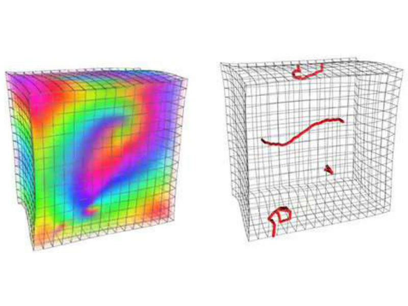
By GlobalDataIn order to visualise the trembling movements inside the heart muscle in three dimensions and to correlate them with the electrical excitation of the heart, the researchers developed new high-resolution ultrasound measuring methods. By analysing image data of the muscle contractions, they were able to observe exactly how areas of contracted and relaxed muscle cells move in a vortex through the myocardium during fibrillation.
They also observed filament-like structures that were previously known to physicists only in theory and from computer simulations. Such a filament-like structure resembles a thread and marks the eye of the whirlpool-like wave or cyclone moving through the myocardium. It is now possible for the first time to pinpoint these centres of the vortices inside the myocardium.
In addition to ultrasound scans, the researchers used high-speed cameras and fluorescent markers that reveal the electrophysiological processes in the myocardium. The images obtained confirmed that the mechanical vortices correspond very well with the electrical vortices.
According to the researchers in Göttingen, ultrasound technology has progressed tremendously in recent years in terms of image quality and imaging speeds, and the potential of modern ultrasound technology has yet to be fully exploited.
Christoph said: “Together with the immensely increased computing power of modern computers and rapid advancements in computer graphics and digital image processing, new measurement and visualization possibilities are being created for investigating the heart. We can apply these developments in medicine today.”
The ultrasound method may also be helpful in the research, diagnosis and treatment of heart failure, during which the myocardial cells do not work effectively as their coordinated contractile movements are disrupted. Doctors would be able to determine the causes with the help of detailed ultrasound scans, enabling them to detect heart failure earlier and treat it more effectively.
