Researchers at the University of Nottingham in the UK have created an ultrasonic imaging system, which can be inserted into the human body for three-dimensional visualisation of cell abnormalities.
Presently in prototype stage, the non-invasive imaging tool called ‘phonon probe’ will be positioned on the tip of a hair-thin optical fibre.
Go deeper with GlobalData

It could be inserted into a standard optical endoscope, a thin tube with a light and camera at the end. This can be steered into the body to detect, assess and operate on cancerous lesions, as well as several other diseases.
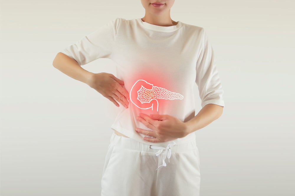
Delivering microscopic and nanoscopic resolution images, the technology can potentially aid doctors in assessing cells in hard-to-reach body parts, such as the gastrointestinal tract. It is also expected to provide better diagnoses for various diseases, including gastric cancer and bacterial meningitis.
Scientists noted that merging optical and phonon technologies could accelerate the clinical workflow process and cut down invasive test procedures for patients.
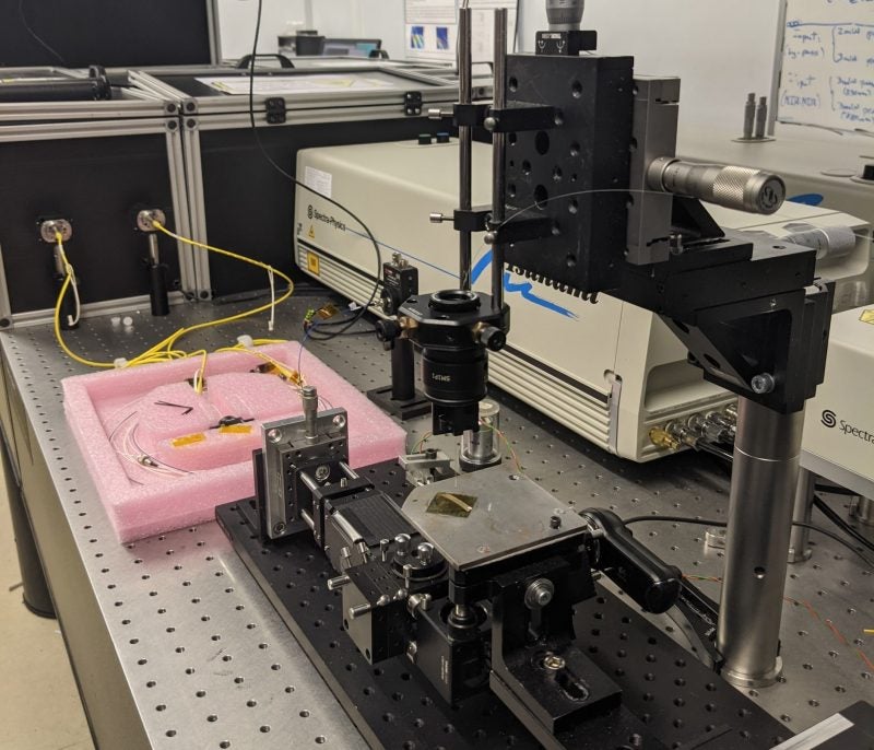
Funded by the Engineering and Physical Sciences Research Council, the phonon probe is a compact system with a performance level that is currently achievable only in research labs with large scientific instruments, the researchers said.
How well do you really know your competitors?
Access the most comprehensive Company Profiles on the market, powered by GlobalData. Save hours of research. Gain competitive edge.

Thank you!
Your download email will arrive shortly
Not ready to buy yet? Download a free sample
We are confident about the unique quality of our Company Profiles. However, we want you to make the most beneficial decision for your business, so we offer a free sample that you can download by submitting the below form
By GlobalDataThe probe can be deployed in clinical settings to enhance patient care and can lower the requirement of standard fluorescent labels.
Fluorescent labels, which are chemicals used for analysing cell biology under a microscope, can harm human cells in high doses.
University of Nottingham Engineering faculty Dr Salvatore La Cavera III said: “We believe the system’s ability to measure the stiffness of a specimen, its bio-compatibility, and its endoscopic potential, all while accessing the nanoscale, are what set it apart.
“These features set the technology up for future measurements inside the body; towards the ultimate goal of minimally invasive point-of-care diagnostics.”
The team now plans to work with the Nottingham Digestive Diseases Centre and the Institute of Biophysics, Imaging and Optical Science to design various biological cell and tissue imaging applications for the development of a viable clinical tool.