
Scientists have developed a new Optical Coherence Tomography (OCT) instrument that will enable users to view the entire eye in unprecedented detail.
Researchers from Nicolaus Copernicus University, Poland, and Universidad de Murcia, Spain, have created a lens that uses an electric current to change its properties, generating higher quality images while avoiding the need for different instruments to look at different parts of the eye. The lens can also view how the vitreous gel that fills the eye interfaces with the retina, which could offer clues on how the retina often becomes detached with ageing.
Go deeper with GlobalData

Discover B2B Marketing That Performs
Combine business intelligence and editorial excellence to reach engaged professionals across 36 leading media platforms.
The research is based on existing OCT imaging systems, which use near-infrared light to create detailed cross-sectional images of the eye. However, traditional imaging systems are limited to imaging depths of two to three millimetres, and struggle to construct a complete image of the eye due to its nature, which bends light to focus on the retina.
The use of an electric current in the instrument, however, has created a dynamic, rather than static, tool for observing the eye.
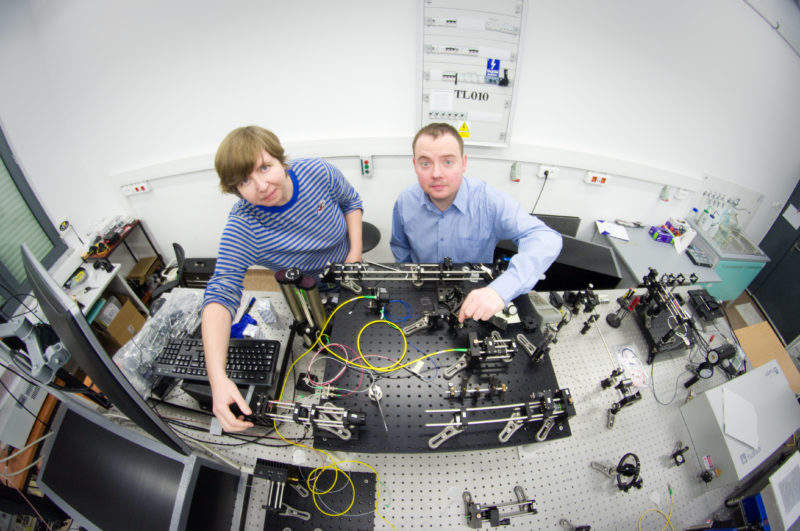
“We incorporated the electrically tuneable lens into a custom-made system that represents the latest generation of OCT technology,” explained Dr Ireneusz Grulkowski, leader of the research team at Nicolaus Copernicus University. “We set out to show that we could image both the front and back of the eye without changing instruments. However, we were also able to show that our instrument enhanced the image quality of the OCT images.”
Grulkowski reported that ‘the study should finish this year’, as the system is complete, but requires approval from the Institutional Review Board. The instrument has several clinical uses, particularly as a tool to identify vitreous detachment.
“We also want to use our instrument to measure opacities in the eye’s critical lens and the vitreous to better understand how various parts of the eye affect the deterioration of vision.” Grulkowski added.

US Tariffs are shifting - will you react or anticipate?
Don’t let policy changes catch you off guard. Stay proactive with real-time data and expert analysis.
By GlobalDataVitreous detachment is a degenerative condition involving the separation of the fibres within the vitreous from the retina that typically affects people aged over 50.
While vitreous detachment itself does not damage sight, the pulling of fibres can lead to the creation of a macular hole in the eye, or a retinal detachment, both of which can lead to permanent loss of vision in the affected eye if untreated.
There are further benefits to the use of a single instrument to observe the eye.
“An instrument that can examine the whole eye will improve the patient’s experience because they won’t have to go through imaging with different devices,” said Grulkowski. “It might also one day reduce the number of instruments – which can be quite expensive – needed in an ophthalmology clinic.”
The team are hopeful that their breakthrough can be used in the future for studies beyond the eye. Grulkowski told me that “we do consider industrial applications of the presented approach. As the first study, we do think that the technology presented in the paper can be used in the inspection device when fast switching between observation planes is required.”
“The demonstrated solution is also useful to increase image quality during imaging the semitransparent materials or the samples, where light penetration is not sufficient.”

