
Researchers from Martin Luther University Halle-Wittenberg (MLU) have developed a new method of generating 3D images of the interior of the human body, which uses laser beams to visualise the development of cancer cells.
The team published its findings in the journal Communication Physics, and describe the technology as a form of photoacoustic imaging. This uses ultrasound waves generated by laser beams to produce high-resolution, 3D images of internal organs.
Discover B2B Marketing That Performs
Combine business intelligence and editorial excellence to reach engaged professionals across 36 leading media platforms.
“Our aim is to visualise cancer cells inside the living body to find out how they function, how they spread and how they react to new therapies,” said MLU medical physicist Professor Jan Laufer. He also identified a key difficulty associated with internal imaging; tumour cells are transparent and so cannot easily be visualised.
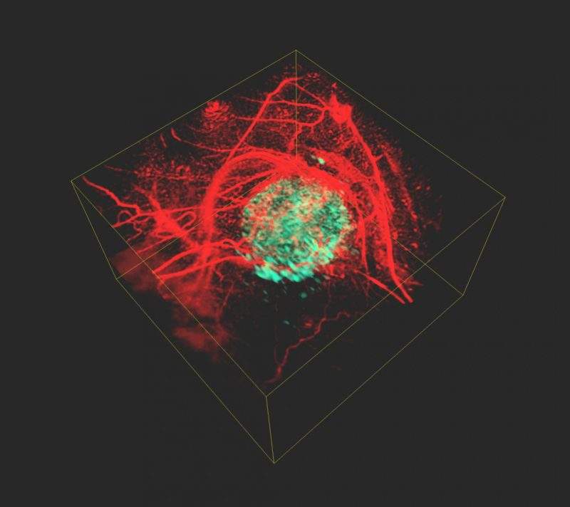
The team’s solution involves introducing a gene into the genome of the cancer cells that produces a phytochrome protein taken from plants and bacteria. This protein operates as what Laufer calls a ‘light sensor’. Next, the team illuminates the tissue with pulses of light at different wavelengths, which are absorbed and converted into ultrasonic waves by the body. These waves can be measured from outside the body, enabling the scientists to reconstruct an image of the body’s insides.
“The special feature of phytochrome proteins is that they alter their structure and thus also their absorption properties, depending on the wavelength of the laser beams,” explained Laufer. “This results in changes to the amplitude of the ultrasound waves that are generated in the tumour cells.
“None of the other tissue components, for example blood vessels, have this property; their signal remains constant,” he added.
The technique produces two different images, enabling scientists to calculate the difference between them and generate a high-resolution, 3D image of the tumour cells. This image of the tumour cells is free of otherwise overwhelming background contrast.
This method could be used to observe cellular and genetic processed in living organisms, beyond identifying cancer cells.
