
New brain-mapping technology could force scientists to rethink how areas of the cerebral cortex communicate.
The technology, called MAPseq, was developed at Cold Spring Habor Laboratory (CSHL) by an international team of scientists led by Professor Anthony Zador. It has allowed the researchers to determine that neurons in the primary visual cortex communicate with higher visual areas of the cortex on a much wider scale than previously thought.
Go deeper with GlobalData

Discover B2B Marketing That Performs
Combine business intelligence and editorial excellence to reach engaged professionals across 36 leading media platforms.
The cerebrum is the largest part of the mammalian brain and contains the cerebral cortex. The wiring diagram of the cortex determines how information, such as sight and sound, is processed across dozens of cortical areas.
Stanford University postdoctoral researcher Jutus Kebschull, who played a key role in developing MAPseq as a graduate student in Zador’s lab, said: “If we don’t know how information is combined even at the earliest stages, then we have essentially no shot of figuring out how the brain works.”
When MAPseg in an experiment, hundreds of thousands of neurons are labelled uniquely with random RNA sequences, which act as barcodes, via a single injection made in the brain. These sequences are transported into the branching axons of each labelled neuron, where they are read out by high-throughput barcode sequencing after the brain is dissected. The process allows scientists to identify every brain region that each barcoded neuron has made contact with.
The CSHL researchers have begun to use MAPseg to compare the brains of a mouse autism model with a healthy mouse model to observe if mis-wiring occurs during development at the single-neuron level, which could explain some of the disorder’s symptoms.

US Tariffs are shifting - will you react or anticipate?
Don’t let policy changes catch you off guard. Stay proactive with real-time data and expert analysis.
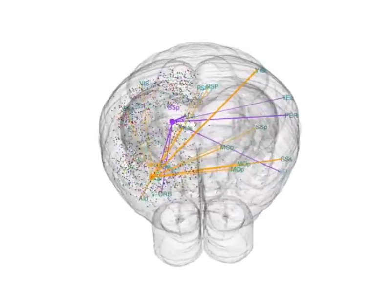
By GlobalDataIn the experiments, the team verified the technology by comparing its results with the reliable mapping method, single-neuron tracing. The researchers used single-neuron tracing to trace 31 mouse neurons in the primary visual cortex to up to seven different cortical areas. Although these experiments took three years to complete, it only took three weeks to use MAPseq to map the projections within the cortex arising from 591 visual cortex neurons.
Kebshull said: “Our finding signals a shift away from the rather convenient idea of every neuron projecting to just one cortical area. That thinking ignores the underlying structure of the brain, and in the future, the way people do their experiments is going to change drastically.”
A video of Zador explaining the new brain mapping technology can be found here: https://www.youtube.com/watch?v=LyWqb9JFUCU#action=share

