An MRI scanning study has revealed that babies with the neurodevelopmental condition fragile X syndrome have less-developed white matter compared with infants without the condition.
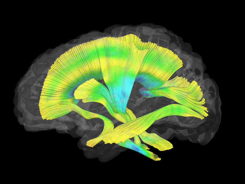
Researchers from the University of North Carolina (UNC) School of Medicine conducted imaging of various sections of white matter in infant brains from different angles to explore the underlying brain circuitry that is integral to healthy neuron communication.
Go deeper with GlobalData

Results from the study show that infants with fragile X syndrome have brain differences related to the disorder already present well before a diagnosis is typically made at the age of three or older.
Heather Hazlett, assistant professor of psychiatry at the UNC School of Medicine, said: “One of the exciting things about our findings is that the white matter differences we observe could be used as an objective marker for treatment effectiveness.”
Previous drug trials have failed to show a change in treatment targets in fragile X patients. One of the challenges for drug developers has been identifying good treatment outcome measures or biomarkers that can highlight intervention responses.
Fragile X syndrome is a genetic disorder and the most common inherited cause of intellectual disability in males. The disorder has several tell-tale symptoms which include learning difficulties, problems with social interaction, delayed speech, hyperactivity and repetitive behaviours. Around 10% of fragile X sufferers have seizures and about 33% meet the diagnostic criteria for autism spectrum disorder.
How well do you really know your competitors?
Access the most comprehensive Company Profiles on the market, powered by GlobalData. Save hours of research. Gain competitive edge.

Thank you!
Your download email will arrive shortly
Not ready to buy yet? Download a free sample
We are confident about the unique quality of our Company Profiles. However, we want you to make the most beneficial decision for your business, so we offer a free sample that you can download by submitting the below form
By GlobalDataMeghan Swanson, co-first author and postdoctoral research fellow at the Carolina Institute for Developmental Disabilities at the UNC School of Medicine, said: “It’s our hope that earlier diagnosis and intervention will help children with fragile X and their families. We also hope that this knowledge might inform drug development research.”
The study involved Swanson, Hazlett and their team using an MRI scanner to scan the brains of 27 infants who went on to be diagnosed with fragile X and 73 who did not develop the disorder. The researchers focused on 19 white matter fibre tracts in the brain as the communication between these bundles of axons is essential to neurodevelopment during childhood.
Analysis of the collected images showed significant differences in the development of 12 of the 19 fibre tracts in infants with fragile X from as early as six months old. The babies who eventually received a positive fragile X diagnosis had significantly less-developed fibre tracts in various parts of the brain.
Jason Wolff, co-first author of the study and assistant professor of educational psychology at the University of Minnesota, said: “These results substantiate what other researchers have shown in rodents—the essential role of fragile X gene expression on early development of white matter in babies.
“Our work highlights that white matter circuitry is a potentially promising and measurable target for early intervention. However, achieving the goal of infant intervention for fragile X would likely require expanded new-born screening efforts.”