
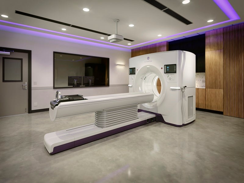
Revolution™ Vibe is a computed tomography (CT) scanner produced by GE HealthCare for head, whole-body, cardiac and vascular X-ray CT applications.
The device was granted 510(k) clearance by the US Food and Drug Administration (FDA) in September 2025.
Recommended White Papers
Recommended Buyers Guides
Featuring Unlimited One-Beat cardiac imaging and AI tools, the scanner is intended to assist clinicians in detecting possible coronary obstructions while improving patient experience and workflow efficiency.
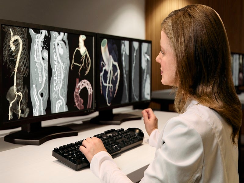
The non-invasive system is designed to produce consistent, high-quality images in cases such as atrial fibrillation and marked coronary calcification.
Revolution Vibe CT System design and features
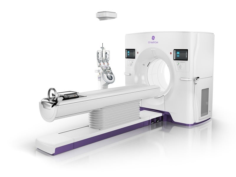
Revolution™ Vibe system includes a gantry, patient table, system cabinet, operator console, power distribution unit and related accessories.
Derived from the Revolution Apex Elite, Revolution™ Vibe is a modified dual-energy CT system.
It retains most of the technologies present in the predicate device, including high tube current output, an 80cm bore with Whisper Drive and deep learning-based image reconstruction for noise reduction.
It also features Adaptive Statistical Iterative Reconstruction (ASIRV), extended field-of-view reconstruction (MaxFOV 2), a rotation speed down to 0.23 seconds per rotation and 160 mm detector span for cardiac examinations.
The primary alteration from the predicate is a revised detector design and associated software adjustments intended for cardiac imaging, which enables full-heart imaging in a single rotation, as with the predicate device.
The detector uses the Gemstone scintillator with configurations of 256 × 0.625mm rows, yielding up to 160mm coverage in the Z axis within a 320mm scan field of view, and 64 × 0.625mm rows, yielding up to 40mm coverage in the Z axis within a 50mm scan field of view.
Revolution Vibe technology and application details
Revolution™ Vibe is designed to support cardiac CT imaging using a single heartbeat acquisition while maintaining diagnostic capability for the rest of the body.
The single beat acquisition is intended to deliver reliable, diagnostically useful images to support clinical decisions, including for technically difficult patients.
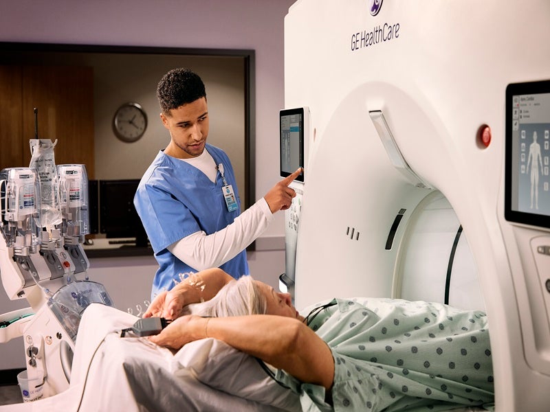
The system integrates an ECG free cardiac option, the True Fidelity DL reconstruction, Snap Shot Freeze 2 motion correction and Effortless Workflow tools that use AI to streamline image acquisition and processing.
The ECG free option can be used in CT studies where an ECG lead is not attached, which may be appropriate when rapid access or simplified setup is required.
TrueFidelity DL uses a deep neural network trained on highquality filtered back projection datasets to distinguish noise from true signal and reduce noise while preserving anatomical and pathological detail.
SnapShot Freeze 2 applies automated wholeheart motion correction, addressing movement of coronary arteries, valves, myocardium, cardiac chambers and major vessels.
Used together, the reconstruction and motioncorrection methods produce cardiac images with reduced motion artefact.
The Effortless Cardiac Workflow utilises AI to choose scan protocols and assist with patient positioning, aiming to reduce scan time and simplify operation.
The system provides spectral imaging via rapid kilovolt (kV) switching and can perform cardiac scans without ECG gating.
Revolution Vibe benefits
The Revolution™ Vibe can produce full heart images at low radiation dose for cases such as atrial fibrillation, patients who struggle to hold their breath, heavily calcified coronary arteries, suspected in stent restenosis, and studies undertaken without an ECG trace.
The system is intended to improve operational efficiency, assist healthcare providers in broadening their services, contribute to better patient outcomes and control lifecycle expenses.
The device has an energy saving design, offers staff training, includes fleet management tools and a Smart Subscription to keep equipment current.
Energy Saving Mode 2.0 switches the unit into a low power state during long idle periods, which lowers daily energy use (kWh) by more than 30%, as per the COCIR SRI 2025 testing method.
A clinical assessment found the one step decision tree workflow cut exam duration by half, saving about four minutes of radiologist time per study.
Patient preparation time was also reduced by as much as five minutes per scan.
These savings were largely due to quicker positioning, less frequent use of beta blockers and avoiding an ECG connection when cardiac exams are prioritised for faster patient access.