
Researchers at Politecnico di Milano, Italy have developed an optical mammography (OM) instrument that uses harmless red or infrared light for diagnostic imaging or monitoring of breast cancer patients.
The device is meant to be used in combination with X-rays to avoid high ionising radiation in people who require repeated imaging. It has been developed under the Smart Optical and Ultrasound Diagnostics of Breast Cancer (SOLUS) project.
Go deeper with GlobalData

Discover B2B Marketing That Performs
Combine business intelligence and editorial excellence to reach engaged professionals across 36 leading media platforms.
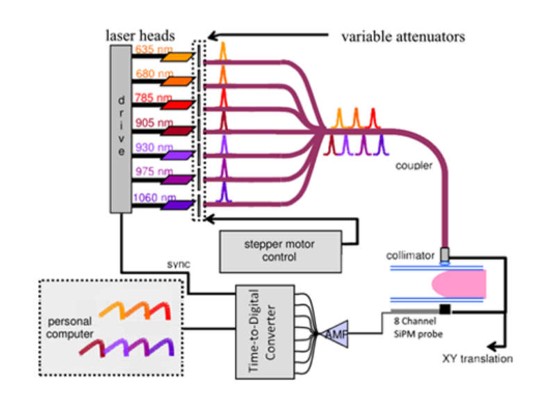
Instead of two photomultiplier tubes (PMTs) seen in current X-ray mammography devices, the new instrument contains an eight-channel probe with silicon photomultipliers (SiPMs) and a multichannel time-to-digital converter.
The latest additions are intended to avoid a time-consuming pre-scan commonly performed to not damage the PMTs, and are said to be responsible for the increased sensitivity and decreased expenses.
Optical imaging methods can be used to analyse tissue composition, including measurement of blood volume, oxygenation, lipid, water and collagen content in a suspected area identified using conventional X-ray imaging technique.
However, OM imaging lacks in the spatial resolution area and its combination with other imaging approaches is expected to address this challenge.

US Tariffs are shifting - will you react or anticipate?
Don’t let policy changes catch you off guard. Stay proactive with real-time data and expert analysis.
By GlobalDataOM instruments also require the exertion of gentle pressure or some need none at all as they work by employing rings of light sources and detectors. OM is considered best applicable in pre-surgical chemotherapy.
Research co-author Edoardo Ferocino said: “This technique is able to provide information on the outcome of chemotherapy just weeks after beginning treatment, or possibly even sooner.”
Ferocino and his team intend to further study the applications of OM in monitoring and predicting chemotherapy outcomes.

