

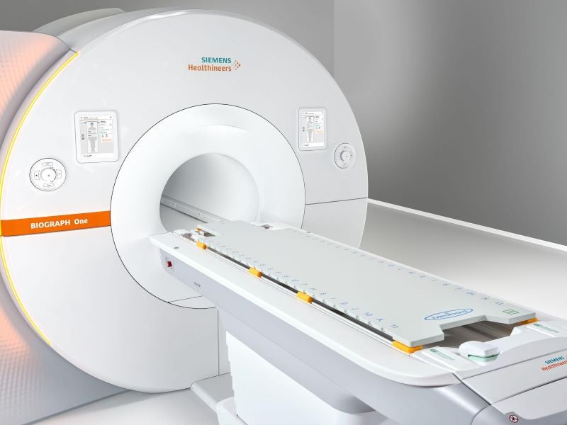
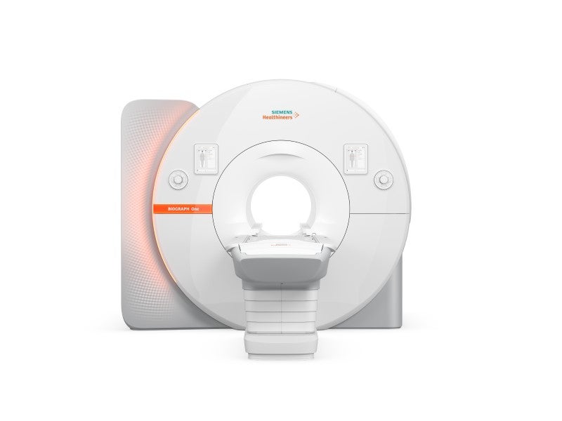
Biograph One is a positron emission tomography (PET) and magnetic resonance (MR) scanner developed by Siemens Healthineers.
The scanner integrates anatomical and functional data acquisition in one scan, supporting the concurrent visualisation of organ position, physiological function and cellular metabolism.
Recommended White Papers

Recommended Buyers Guides

The system was first presented at the Radiological Society of North America 2024 meeting, held in December 2024.
Biograph One obtained the CE Mark in Europe in September 2025 and subsequently received 510(k) clearance from the US Food and Drug Administration in January 2026.
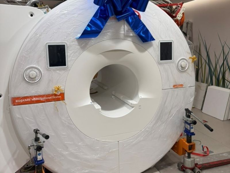
Rigshospitalet in Denmark is the first hospital worldwide to install and conduct clinical investigations with the PET/MR system, followed by the University of Iowa in the US.
Biograph One design and features
Biograph One is a second-generation scanner that builds on Siemens Healthineers’ first commercial PET/MR platform, the Biograph mMR, which was introduced in 2011.
Its MR component is derived from the Magnetom Vida 3 Tesla scanner, while its Optiso UDR Pro digital PET detector, which uses lutetium oxyorthosilicate crystals, is based on the Biograph Vision PET/CT system.
The system features an open reconstruction architecture and is equipped with a 3T magnet, which offers a 55cm³ × 55cm³ × 50cm³ field-of-view (FOV). It is engineered for high B0 homogeneity and high gradient power to support demanding clinical and research protocols.
The 35cm axial FOV digital PET detector of the scanner is described as the largest axial FOV currently available in PET/MR. The detector supports the imaging of larger anatomical regions in a single examination and provides a time-of-flight (TOF) performance of 187 picoseconds under increased gradient loads.
The PET detector, together with the 3T magnet, supports the visualisation of small lesions and high soft-tissue contrast.
The scanner also incorporates new, flexible BioMatrix Contour XL MR coils, designed in a blanket-like format to provide whole-body coverage with fewer coils, which can reduce setup time and limit technologists’ exposure to radiation.
Technology details
The Biograph One features the Deep Resolve Boost 2D and 3D reconstruction technology, which is intended to enable faster acquisition and improved spatial resolution in both 2D and 3D PET/MR applications, while reducing noise.
An integrated BioMatrix Position Sensor communicates with the system to recommend suitable patient positioning, with the aim of reducing examination preparation time.
The scanner uses the MyExam Companion user interface, which offers advanced automation tools intended to streamline the entire scanning workflow for radiologic technologists.
Biograph One offers whole-body scanning modes, myExam SinglePass and MultiPass, to provide configurable acquisition options that can be adapted to individual examination requirements.
It also incorporates workflow tools aimed at simplifying patient positioning and examination setup, enabling operators to initiate scans directly at the scanner and use BioMatrix Contour coils to further streamline procedures and reduce time spent on preparation.
Additionally, advanced AI-driven algorithms speed up the scanning process while enhancing overall image quality.
Biograph One benefits
The Biograph One device combines quantitative PET performance, ultra-fast TOF capability and high-quality MR imaging within a single platform, supporting patient-focused imaging pathways.
The large PET detector is designed to reduce whole-body acquisition time by requiring fewer bed positions, with reported scan durations of under 30 minutes to align more closely with typical radiology scheduling and to accommodate higher patient throughput.
In oncology, the wider coverage enables multiple disease sites to be assessed in a single session; for instance, imaging brain tumours alongside lymph node metastases.
By acquiring PET and MR data within a single integrated examination, the system is designed to support clinical decision-making across multiple stages of care, from initial diagnosis and staging through therapy selection and follow-up assessments.
This integration creates opportunities for theranostics by bringing diagnostic assessment and treatment delivery into closer alignment, supporting more personalised care. In Alzheimer’s disease, clinical trials suggest that the combination of PET/MR could evolve into a standard, single-visit imaging solution for routine clinical use.
