
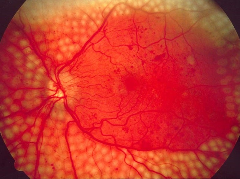
Research from the Joslin Diabetes Center’s Beetham Eye Institute in Boston, Massachusetts has shown that ultra-wide field (UWF) fluorescein angiography detects over three times more microaneurysms than UWF colour imaging, suggesting that the two techniques should not be used interchangeably.
UWF imaging allows clinicians to see over 80% of the retina in a single image, with UWF colour imaging used more frequently in the US as it is non-invasive.
Go deeper with GlobalData

Discover B2B Marketing That Performs
Combine business intelligence and editorial excellence to reach engaged professionals across 36 leading media platforms.
Diabetic retinopathy is diagnosed and graded using UWF imaging, as determining the number and location of microaneurysms is key to determining the risk of diabetic retinopathy progression.
Clinicians have historically used fluorescein angiography and colour imaging interchangeably when assessing the condition.
Joslin Diabetes Center chief retina fellow Dr Mohamed Elmasry said: “Existing research often uses fluorescein angiography and colour imaging as two interchangeable sides of the same coin when assessing microaneurysms. However, our research shows that these modalities aren’t the same and we have to be cautious before using them in that way.”
A total of 193 patients with diabetic retinopathy had UWF colour imaging and UWF fluorescein angiography done on the same day. In total, 288 eyes were included in the analysis.
Researchers manually counted microaneurysms from both imaging techniques and directly compared the results. They compared the number of microaneurysms they identified at each level of disease severity and compared those results.
Overall, fluorescein angiography detected 3.5 times more microaneurysms than colour imaging, even after researchers adjusted for the patient’s diabetes duration, average blood sugar levels and gender.
Fluorescein angiography also identified up to 3.5 times more retinal areas with more than 20 microaneurysms, an indication of severe retinopathy, than colour imaging.
However, when a correction factor of four was applied to the fluorescein angiography results for each grade of disease severity, the techniques did become more comparable.
Beetham Eye Institute director Dr Lloyd Aiello said: “This study should help inform artificial intelligence and other imaging studies looking to automate the grading process using ultra-wide field colour imaging or fluorescein angiogram, and make them more precise and more comparable.”
