
A team of Google researchers has developed a tool that combines augmented reality (AR) with a deep learning neural network that can help pathologists to spot cancerous cells on slides under a microscope.
The researchers described their augmented reality microscope (ARM) platform in a paper published on the company’s website and gave a demonstration of the device at this year’s American Association for Cancer Research meeting.
Go deeper with GlobalData

Discover B2B Marketing That Performs
Combine business intelligence and editorial excellence to reach engaged professionals across 36 leading media platforms.
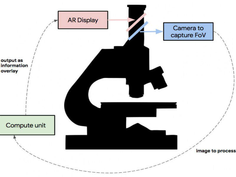
The ARM platform works by highlighting potentially cancerous cells with a red ring that alerts the pathologist carrying out a biopsy to take a closer look.
Examining a biopsy sample of cancerous tissue can be an intense process. The thinly sliced sample is viewed on a glass slide under a microscope by a pathologist trained to look for cancerous cells.
The Google team aims to make this job easier as it can be tedious and lead to fatigue and errors. To do this, the researchers built on previous work at the company, which involved a team developing a deep learning technique that could spot breast cancer.
The researchers incorporated lessons learned from various AR projects to train their deep learning application to draw circles around suspected cancer cells in the tissue samples. Their system was interfaced with the most commonly used pathologist microscope so that the tool can just be added and pathologists will not be required to get a new version.
Google has reported that the platform is now capable of assisting with the detection of breast and prostate cancer. In the future, the company claims the tool could be trained to spot other cancers and some types of infectious diseases such as TB and malaria. It could also be programmed with other AR capabilities, including the ability to post labels over tissue sections, while providing arrows or other indicators that could aid pathologists.
A further explanation of the device can be found here: https://www.youtube.com/watch?v=9Mz84cwVmS0#action=share
