
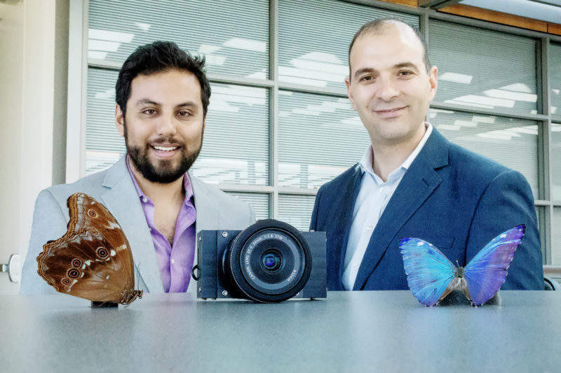
Researchers have developed a surgical camera inspired by the eyes of the morpho butterfly, which perceives infrared signals given off by dyes that bind to tumours in the body, enabling doctors to locate and remove all of a patient’s cancerous tissue.
The team, from the University of Illinois at Urbana-Champaign and Washington University in St. Louis published their findings in the paper ‘A bio-inspired imager improves sensitivity for near-infrared fluorescence image-guided surgery’ in the journal Optica.
Go deeper with GlobalData

Discover B2B Marketing That Performs
Combine business intelligence and editorial excellence to reach engaged professionals across 36 leading media platforms.
“By looking at the way nature has designed the visual systems of insects, we can address serious problems that exist with cancer surgery today and make sure there are no cancer cells left behind during surgery,” said Viktor Gruev, Illinois professor of electrical and computer engineering and of the Carle Illinois College of Medicine, who led the study.
“This technology is more sensitive, more accurate, much smaller and lower-cost than currently available instruments that are FDA-approved to detect these signals.”
Gruev and his team built the camera with the same nanostructures that are found in the eyes of a morpho butterfly, which can see multispectral images, including near-infrared. This enables the camera to simultaneously register colour images and near-infrared signals without having to change external lighting. The camera is connected to surgical goggles, to make the system easy to use.
“The surgeon puts on the goggles that have integrated our bio-inspired camera technology, and it will protect their eyes and at the same time project the fluorescent information whenever they want it,” said Gruev.
“The goggles are also incredibly low-cost. We anticipate it to cost around $200, compared with $20,000 for the cheapest FDA-approved instrument.”
The camera was tested on mice and humans, and was able to identity a near-infrared fluorescent dye that binds to the breast cancer cells within the mice. The surgeons were also able to locate tumour sites without penetrating the skin, as the camera can pick up signals from beneath the surface of the tissue.
The researchers also used a green dye to highlight lymph nodes in human patients with breast cancer, as the removal of lymph nodes around a tumour will reveal if the tumour has spread. The camera identified cancerous lymph nodes in two patients that the surgeons did not see visually.
The camera addresses the existing problems of identifying cancerous tissue; many hospitals use near-infrared fluorescent agents that bind to tumours and can be seen through specialised displays, but the necessary machines are costly and can only function in dimmed light, making it difficult for surgeons to see.
The team has filed for a patent for the camera technology, and will then work to integrate their camera with endoscopic camera systems, enabling surgeons to have a guided first-person view of a patient’s insides.

