
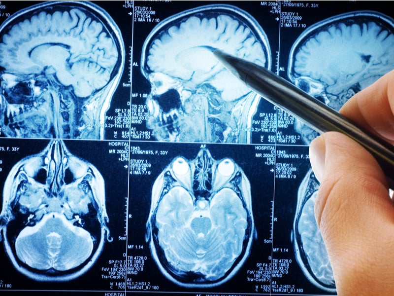
Two studies presented at the annual meeting of the Radiological Society of North America (RSNA) have used magnetic resonance imaging (MRI) to illuminate abnormalities in the brains of people with depression.
Researchers at Stony Brooke University Renaissance School of Medicine have recently studied the connection between depression and disruptions to the blood brain barrier (BBB), a network of blood vessels and tissue which protects the brain from foreign substances.
Go deeper with GlobalData

Discover B2B Marketing That Performs
Combine business intelligence and editorial excellence to reach engaged professionals across 36 leading media platforms.
Using an MRI technique known as intrinsic diffusivity encoding of arterial labelled spins (IDEALS), they looked at BBB water permeability, the movement of water out of the blood vessels and into the brain tissue.
Comparing 14 healthy individuals and 14 patients with depression, the researchers found that less water moved from inside the blood vessels to outside in depressed patients.
This disrupted BBB integrity was especially prominent in the amygdala and the hippocampus, regions of the brain known to be altered in major depressive disorder.
Columbia University researcher and co-author of the study Dr Kenneth Wengler said: “This study helps improve our understanding of the pathophysiology of depression and can open new avenues of treatment for a disorder that affects over 100 million individuals worldwide.”
A second depression MRI study presented at the 2019 meeting looked at abnormalities in the connectome of the brain and their role in depression. Researchers at the University of North Carolina (UNC) compared 66 adults with depression with 66 matched controls during wakeful rest using functional MRI (fMRI) and a newly developed multiscale neural model inversion framework that linked the brain’s microscopic circuitry with its larger-scale interactions.
Researchers were able to use these scans to interpret the excitatory or inhibitory influence between neuronal cell groups, a proper balance between which is crucial for a well-functioning brain. Depressed patients were found to have abnormal patterns of both excitation and inhibition at the dorsal lateral prefrontal cortex, an area of the brain vital to cognitive control functions including the regulation of the amygdala.
The researchers found that recurrent excitation in the thalamus, an area of the central brain that is also responsible for emotional regulation, was abnormally elevated in patients with depression.
UNC researcher Dr Guoshi Li said: “Current methods of studying the brain provide a superficial understanding of connectivity. This [MRI] method allows us to identify impaired connectivity within each brain region, making it a potentially more powerful tool to study the neuromechanism of brain disorders and develop more effective diagnosis and treatment.”
Depression is one of the most common mental disorders worldwide. Researchers hope a more sophisticated understanding of brain changes associated with the disorder will lead to the development of more effective treatments.
