
A study by NYU School of Medicine, US, has resulted in the development of a new glove shaped MRI component which has delivered the first clear images of bones, tendons and ligaments moving together in a human hand.
The study authors believe the MRI glove has the potential to be useful in the future diagnosis of repetitive strain injuries such as carpal tunnel syndrome. As the device shows how different tissue types impact on each other when they move, it could be used to create a more in-depth atlas of hand anatomy, aid hand surgery with more realistic hand images, or help in the design of better prosthetics.
Go deeper with GlobalData

Discover B2B Marketing That Performs
Combine business intelligence and editorial excellence to reach engaged professionals across 36 leading media platforms.
NYU Langone Health research scientist and lead author Bei Zhang said: “Our results represent the first demonstration of an MRI technology that is both flexible and sensitive enough to capture the complexity of soft-tissue mechanics in the hand.”
MRI works by immersing tissues in a magnetic field so that any hydrogen atoms present align to create an average magnetic force in one direction in each tissue slice. These ‘little magnets’ can be tipped out of equilibrium by radio waves. Once tipped, they spin and emit radio signals which then reveals their positions so that an image can be rebuilt.
Also fundamental to MRI is radiofrequency coils being able to convert radio waves into a detectable electric current. However, this means that the captured spinning radio waves produce little currents inside receiver coils and create their own magnetic fields, which prevents nearby coils from capturing clean signals.
Due to research over the past few decades, MRI scanners have been developed that contain carefully positioned receiver coils, arranged to cancel out magnetic fields in neighbouring coils. Once the best arrangement has been set, coils can no longer move relative to one another, which limits the ability of MRI to image complex moving joints.
The leap made by the study authors was to design a ‘high impedance’ structure that blocks current, and then measures how hard the force in magnetic waves ‘pushes’ the voltage as it attempts to create a current in the coil.
As no electric current is created by the MR signal, the new receiver coils no longer create magnetic fields that interfere with neighbouring receivers. This removes the need for rigid structures.
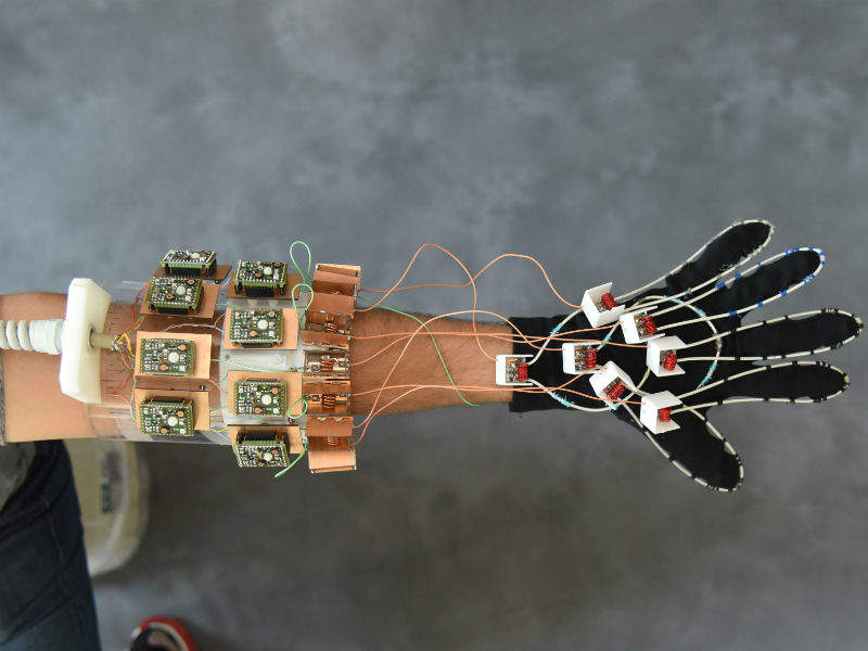
The researchers claim that their system, with coils stitched into a cotton glove, generated ‘exquisite’ images of freely moving muscles, tendons and ligaments in a hand as it played the piano and grabbed objects.
The MRI signal is produced by protons and so excels at imaging soft tissue structures rich in water because each water molecule includes two atoms of hydrogen. Therefore, MRI is good at imaging muscles, nerves, and cartilage, which are difficult to study using other non-invasive methods. However, tendons and ligaments remain difficult to see independently, because both appear as black bands running alongside bone.
The study found that, when visualising flexing fingers, the new coils revealed how the black bands moved with the bones, which could help to record differences that come with injury.
NYU Langone Health assistant professor and senior author Martijn Cloos said: “We wanted to try our new elements in an application that could never be done with traditional coils, and settled on an attempt to capture images with a glove.
“We hope that this result ushers in a new era of MRI design, perhaps including flexible sleeve arrays around injured knees, or comfy beanies to study the developing brains of newborns.”
