
A team of professionals led by the University of Cambridge has developed a new algorithm to monitor the joints of arthritis patients and track small changes to enable a better understanding of the condition.
The new technique is expected to help in determining the effectiveness of the new treatments on osteoarthritis without invasive tissue sampling.
Go deeper with GlobalData

Discover B2B Marketing That Performs
Combine business intelligence and editorial excellence to reach engaged professionals across 36 leading media platforms.
Osteoarthritis is one of the most common forms of arthritis in the UK and is caused when articular cartilage wears out, resulting in immobile and painful joints.
It is generally identified through an x-ray when the space between two bones at the joint narrows down. However, an x-ray cannot help to detect minute changes at the joint.
Cambridge Department of Engineering official Dr Tom Turmezei said: “In addition to their lack of sensitivity, two-dimensional x-rays rely on humans to interpret them.
“Our ability to detect structural changes to identify disease early, monitor progression and predict treatment response is frustratingly limited by this.”
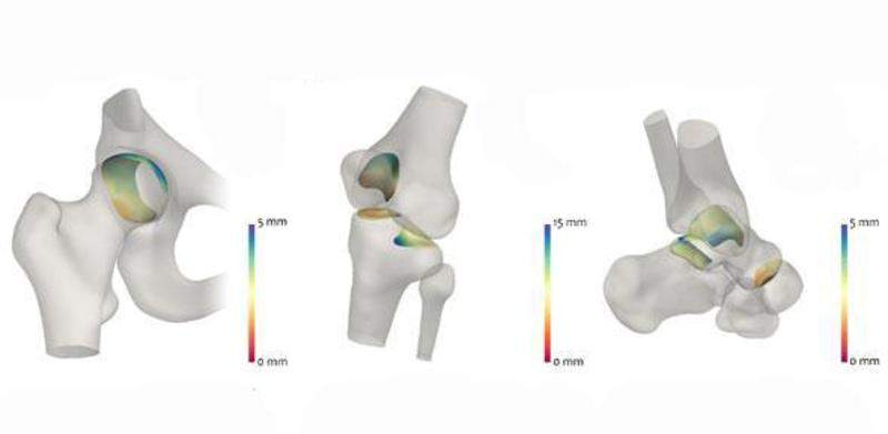
Under the new joint space mapping (JSM) technique, the images from a standard computerised tomography (CT) scan are used to receive detailed images of the affected joints in 3D.
Subsequently, these CT images are analysed to detect the changes in the space between the bones of the respective joint.
An algorithm is generated which includes tests on human hip joints from bodies donated for medical research. The researchers found that it was twice as good at identifying small structural changes compared to the conventional process.
The JSM algorithm produces colour-coded images identifying the areas where the space between the bones has changed.
Turmezei added: “Using this technique, we’ll hopefully be able to identify osteoarthritis earlier and look at potential treatments before it becomes debilitating.
“It could be used to screen at-risk populations, such as those with known arthritis, previous joint injury, or elite athletes who are at risk of developing arthritis due to the continued strain placed on their joints.”
