

US researchers at the University of California, Los Angeles (UCLA) have developed a new system to aid easy and less expensive diagnosis of chronic diseases in remote areas.
Designed to create three-dimensional (3D) images of tissue samples, the new system features optical hardware, a lens-free microscope, and algorithms.
Go deeper with GlobalData

Discover B2B Marketing That Performs
Combine business intelligence and editorial excellence to reach engaged professionals across 36 leading media platforms.
The system is intended to replace costly lab equipment primarily in developing countries and remote areas that do not have access to the existing devices for tissue biopsies.
It can be used to make biological samples transparent and later image them using a lens-free microscope.
UCLA Electrical and Computer Engineering and Bioengineering professor Aydogan Ozcan said: “Although technological advances have allowed physicians to remotely access medical data to perform diagnoses, there is still an urgent need for a reliable, inexpensive means for disease imaging and identification, particularly in low-resource settings, for pathology, biomedical research and related applications.”
To prepare the samples, the research team used a technique called Clarity that employs a chemical process to remove fat and retain proteins and DNA in the tissue, making it transparent.
To stain the samples, the team used coloured, light-absorbing dyes, which did not have noticeable signal degradation over time.
The researchers then used their newly developed holographic lens-free microscope device to produce 3D images using one-tenth of the image data required by standard scanning optical microscopes.
Thick samples and large sample volumes can be studied using the new device, providing an advantage during conditions where equipment for obtaining thin samples is not accessible.
The researchers used a silicon chip with millions of photo detectors for mounting the tissue sample to allow enhancement of resolution and view of various cross-sections of the sample.
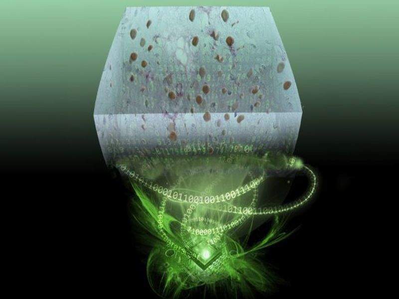
Image: Rendering of a lens-free holographic microscope that uses a silicon chip and computer algorithms to create 3-D images of tissue samples. Photo: courtesy of Ozcan Research Group/UCLA.

