
A research team at the University of Nottingham has developed an endoscopic probe enabling practitioners to three-dimensionally (3D) image the stiffness of individual biological cells and complex organisms to discover and treat cancer earlier.
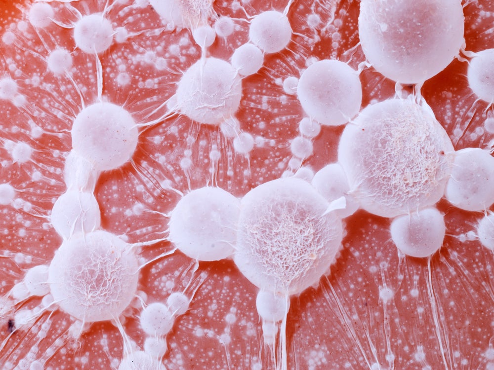
Cancer cells are softer than regular cells in their early stages, which enables them to squeeze through tight gaps and metastasise throughout the body. During this process, cancer cells modify their surrounding environment and create stiff tumours, which insulate them from outside threats.
Go deeper with GlobalData

Discover B2B Marketing That Performs
Combine business intelligence and editorial excellence to reach engaged professionals across 36 leading media platforms.
By measuring the stiffness of individual cells, the university’s optics and photonics group said the device will be able to evaluate microscopic cellular tissue based on abnormal stiffness at the single cell level inside the human body for the first time.
The device, which follows the engineering faculty at Nottingham University’s development of an ultrasonic imaging system for 3D visualisation of cell abnormalities in 2021, achieves high imaging resolution to detect the stiffness of objects down to billionths of a metre (nanometres) through a physical phenomenon known as Brillouin scattering, in which a laser beam interacts with the natural stiffness of a specimen.
To demonstrate this capability, the team visualised the 3D stiffness of Caenorhabditis elegans (C. elegans), a microscopic organism known scientifically as a nematode.
The high imaging of the probe helped provide visualisation and material information about the nematode’s cuticle, a part of the organism’s anatomy which has reportedly only previously been imaged through electron microscopes in non-living conditions.
Dr Salvatore la Cavera, lead research author and Nottingham research fellow in the optics and photonics group at the University of Nottingham commented: “Our device makes it possible to ‘feel for a stiff lump,’ but on a single cellular scale, meaning we could catch cancer early at microscopic cell scales rather than large malignant tumour scale.
“It is non-invasive, non-toxic, and very promisingly, is related to technology that can quantitively determine the presence of cancer cells using artificial intelligence – providing a chronically understaffed area with a much-needed solution to a real-world problem that the industry has faced for decades.”
On future developments for the device, la Cavera told Medical Device Network there are several avenues the team are aiming at in the development of imaging and sensing applications.
“We will develop a fibre imaging bundle version of this technology to enable microscopic stiffness imaging down the utility port of standard clinical endoscopes; and we will continue to develop an in-vitro imaging system, similar to the one we’ve used for our C. elegans studies that will have improved lateral resolution, imaging depth, and acquisition speed.
“For sensing applications, we will integrate our device into hypodermic and biopsy needles to monitor the stiffness of tissue in real-time during future clinical and surgical procedures.”
The full paper on the team’s research has been released in Communications Biology, a journal published by Nature.
The Food and Drug Administration (FDA) recently approved a related endoscopic device developed by Fujifilm Corp for early-stage colorectal cancer screening.
Fujifilm’s CAD EYE is an artificial intelligence (AI) detection system designed for endoscopic imaging with the aim to enhance the detection of colonic mucosal lesions during colonoscopy procedures and aid in the identification and removal of pre-cancerous lesions.
