
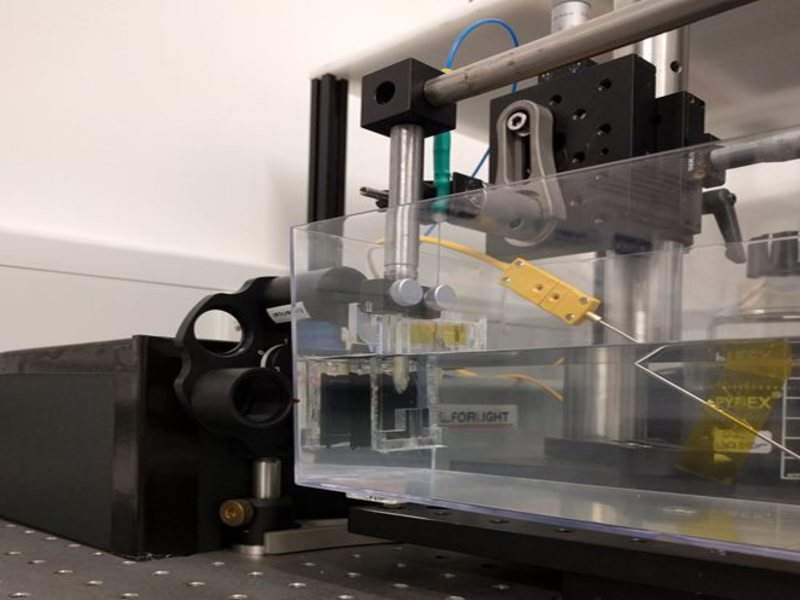
Researchers at University College London, UK, have developed an optical ultrasound system with the potential to improve medical imaging performance for the diagnosis and treatment of health conditions.
The all-optical device eliminates electrical components and allows the safe usage of magnetic resonance imaging (MRI) scanners to gain a better image of the required tissues.
Go deeper with GlobalData

Discover B2B Marketing That Performs
Combine business intelligence and editorial excellence to reach engaged professionals across 36 leading media platforms.
In addition, the new optical ultrasound system integrates lightbeam scanning mirrors to enhance image quality and enable picture acquisition in various modes.
It combines optical ultrasound generating materials, ultrasound source geometries and a fibre-optic ultrasound detector to acquire and display images at video rates.
University College London researcher Erwin Alles said: “The flexibility offered by the scanning mirrors will allow for seamless switching between 2D and 3D imaging, as well as a dynamically adjustable trade-off between image resolution and penetration depth, without the need to swap imaging probe.
“Especially in a minimally invasive interventional setting, swapping imaging probes is highly disruptive, extends procedure times and introduces risks to the patient.”
Unlike standard ultrasound imagers, which use electronic inducers to transmit high-frequency sound waves and receive their reflections, all-optical imagers utilise light to transmit and receive ultrasound waves.
Optical components can also be easily miniaturised and small all-optical ultrasound probes are expected to be significantly less expensive compared to the compact electronic ultrasound systems.
The prototype of the new optical ultrasound system was evaluated through imaging of a deceased zebrafish and a pig artery that was altered to mimic the dynamics of pulsing blood.
Data revealed the imaging capabilities of the new system comparable to an electronic high-frequency ultrasound device. Findings from the research were published in The Optical Society (OSA) Biomedical Optics Express journal.
Currently, the researchers are developing a long, flexible imaging probe for free-hand operation during clinical use, along with miniaturised variants for endoscopic applications.
