
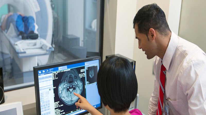
Scientists at Case Western Reserve University in the US have developed a new computer imaging method to enable cancer diagnosis using routine CAT scans.
The new approach is expected to offer a non-invasive, easy, accurate and cost-effective option for determining if certain tumours are cancerous or not.
Go deeper with GlobalData

Discover B2B Marketing That Performs
Combine business intelligence and editorial excellence to reach engaged professionals across 36 leading media platforms.
Lung-cancer screenings typically involve interpretation of suspicious nodules on a CAT scan by a radiologist.
Patients have to then undergo invasive and costly tests such as surgical biopsies for the nodule assessment.
Case Western’s new technique analyses the regions outside the tumour and the surrounding blood vessels to identify whether the nodules are cancerous. This avoids deeper examination or cutting.
Case School of Engineering biomedical engineering professor Anant Madabhushi said: “We’re now doing something that radiologists do not typically tend to do.
“Radiologists have been looking at CAT scans for 40 or 50 years, but they have never looked in these locations or found what we’ve found.”
The new approach uses ‘deep-learning’ diagnostic computers to evaluate the shape of blood vessels feeding a nodule, as well as those in regions near the tumour.
It can accurately predict a cancerous growth by looking for any previously unrecorded changes in these blood vessels or otherwise healthy surrounding tissue.
Researchers believe that this type of diagnosis from an existing CAT scan will enable clinicians to better determine if a patient requires additional invasive procedures.
Madabhushi added: “A lot of these nodules show up on a scan as ‘indeterminate,’ but 98% turn out to be benign, but only after more tests are done. So there’s the economics of it, but also the anxiety for the patient.
“We don’t really do a great job of lung-cancer screening because we don’t have better tools. This would be a far, far better tool.”
During testing, the team demonstrated that the computer technique could successfully analyse the regions outside the tumour and the blood vessels, and predict whether the nodules found on a lung CAT scan are cancerous or harmless.
Findings from the research were published in the Nature and Radiology journals.
