
Bioengineers from Harvard University in the US have developed a 3D model of the human heart’s left ventricle to carry out cardiac disease research.
The researchers seeded a nanofiber scaffold, which acts as a 3D template, with human heart cells. This scaffold guides the cells and their assembly into ventricle chambers.
Go deeper with GlobalData

Discover B2B Marketing That Performs
Combine business intelligence and editorial excellence to reach engaged professionals across 36 leading media platforms.
As the chambers can beat in-vitro, researchers will be able to evaluate heart function by using common clinical tools such as pressure-volume loops and ultrasound.
The team expects the 3D model to help in the study of disorders, drug assessment and developing patient-specific therapies for heart conditions.
Harvard John A. Paulson School of Engineering and Applied Sciences postdoctoral fellow Luke MacQueen said: “The long-term objective of this project is to replace or supplement animal models with human models and especially patient-specific human models.
“In the future, patient stem cells could be collected and used to build tissue models that replicate some of the features of their whole organ.”
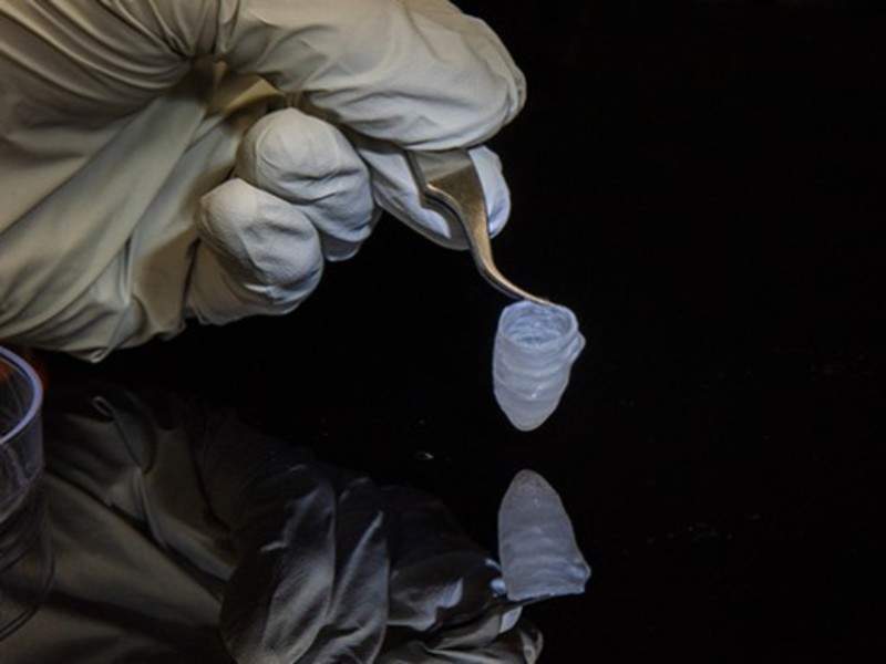
The researchers used a nanofiber production platform called pull spinning to build the scaffold, and combined biodegradable polyester with gelatin fibres to create the ventricle.
Rat myocytes or human cardiomyocytes from induced stem cells were utilised to culture the ventricle. The human cells were able to survive for six months.
Also, the researchers were able to control and track calcium propagation in the beating ventricle, and could insert a catheter for monitoring its pressure and volume.
Furthermore, it was observed that the tissue’s beat-rate increased upon exposure to a drug called isoproterenol, which is similar to adrenaline that causes such reactions in humans and rats.
The team additionally poked holes in the ventricle to imitate myocardial infarction and studied the effect of the resulting heart attack in a petri dish.
Currently, the researchers are planning to utilise patient-derived, pre-differentiated stem cells to seed the ventricles in order to obtain high-throughput production.
