
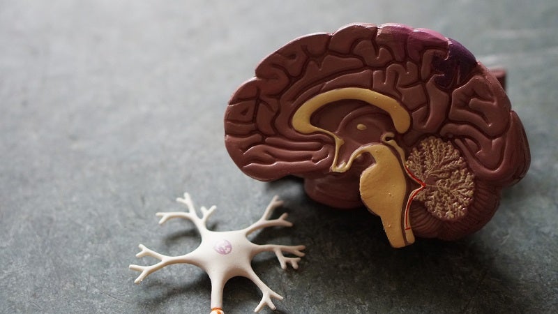
Researchers at the Yale School of Medicine have identified biomarkers of attention-deficit/hyperactivity disorder (ADHD) by analysing the data from magnetic resonance imaging (MRI) exams on approximately 8,000 children.
They have also identified the role of neuroimaging machine learning to assist in diagnosing, treating and observing the disorder.
Go deeper with GlobalData

Discover B2B Marketing That Performs
Combine business intelligence and editorial excellence to reach engaged professionals across 36 leading media platforms.
The findings from the new study are planned to be presented at the Radiological Society of North America’s (RSNA) next annual meeting,
The researchers used MRI data obtained from the Adolescent Brain Cognitive Development (ABCD) study of brain development and child health, which was conducted in the US.
This study involved 11,878 children aged between nine and ten years, of which 7,805 patients, including 1,798 with an ADHD diagnosis, were included in the Yale School of Medicine study group, following exclusions.
All the participants in the trial had structural MRI scans, diffusion tensor imaging, and resting-state functional MRIs taken.
The researchers statistically analysed the imaging data of the participants to determine the relation of ADHD with neuroimaging metrics that included white matter integrity, brain volume, surface area, and functional connectivity.
They found a thinning of the brain cortex and significant white matter microstructural changes, which were mainly in the frontal lobe of the brain in ADHD patients.
Additionally, the researchers observed abnormal connectivity in the brain networks that are involved in memory and auditory processing.
Yale School of Medicine researcher and study co-author Huang Lin said: “We found changes in almost all the regions of the brain we investigated.
“The pervasiveness throughout the whole brain was surprising since many prior studies have identified changes in selective regions of the brain.
“The frontal lobe is the area of the brain involved in governing impulsivity and attention or lack thereof – two of the leading symptoms of ADHD.”
