
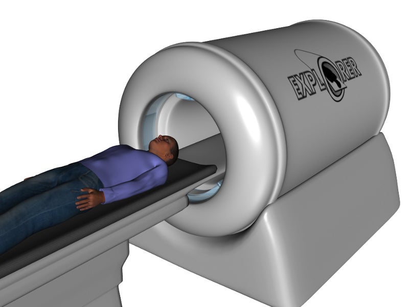
EXPLORER is a positron emission tomography (PET) scanner that produces three-dimensional (3D) pictures of the entire human body.
The scanner was designed in 2017 by engineers and researchers from multiple institutions, including UC Davis Biomedical Engineering, the University of Pennsylvania and the US Department of Energy’s Lawrence Berkeley National Laboratory.
Recommended Buyers Guides
The first EXPLORER PET scanner was manufactured in May 2018 and produced its first images in November that year. It will be installed in early 2019 for research applications at the UC Davis Medical Centre in Sacramento, California.
Data produced by the scanner will be used to extract detailed information regarding metabolism, radiotracer transport, binding and tissues in the whole body.
Development and financing of the EXPLORER total-body scanner
In September 2015, the PET scanner’s development received five-year $15.5m funding from the National Institute of Health (NIH) director’s Transformative Research Award.
The project also received substantial financial support for initial research and electronics development from the National Cancer Institute (NCI) and the National Institute of Biomedical Imaging and Bioengineering (NIBIB).
The feasibility study of a PET scanner for non-human primates was completed in November 2016 and a mock-up of the scanner was delivered to UC Davis the following month.
A prototype of the mini-EXPLORER PET scanner was developed for animal research by UC Davis in partnership with Siemens Medical Solutions.
In February 2018, the mini-EXPLORER II animal PET/CT scanner was installed at the UC Davis School of Veterinary Medicine for scanning animals.
Contractors involved in device development
In January 2017, the EXPLORER total-body PET scanner project consortium awarded a contract to two industry partners, United Imaging Healthcare (UIH) America and SensL Technologies, to fabricate the scanner for humans.
UIH was responsible for building the scanner, while SensL supplied the detectors.
EXPLORER scanner design and features
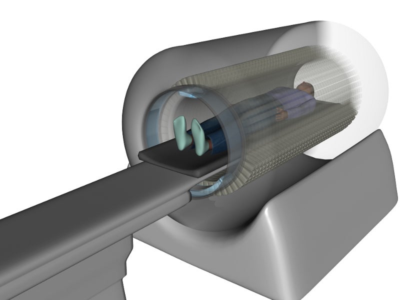
The cylindrical EXPLORER total-body scanner combines PET and X-ray computed tomography (CT) imaging technologies to produce images with high temporal resolution.
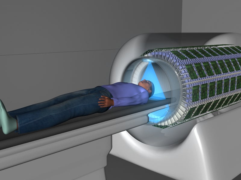
A total of 40 rings and 48 modular block detectors are present in the scanner. The ring diameter is around 80cm and the spatial resolution is 4mm. The device has an acceptance angle of 46°.
The scanner has an axial field of view of 194cm and provides PET images through around 500,000 detector elements.
It is claimed to perform total-body scanning up to 40 times better than existing commercial scanners, performing 3D scans of the whole body within 30 seconds. Its fast-scanning ability avoids the need for anaesthesia when scanning young children.
In addition, the unit incorporates modern solid-state silicon photomultiplier light sensors to provide high-resolution images.
Applications of the total-body scanner
The EXPLORER scanner can be used in a wide range of applications, including drug development, toxicology and the detection of infections and chronic diseases such as cancer.
It provides the ability to identify the pharmacokinetics (pk) of new medicines in all organs of the body at lower masses and radiation doses of 100µSv.
The scanner can also be employed to examine trafficking behaviours in cell therapies, autoimmune disease, metabolic disorders and other chronic conditions. It can also be used in biomedical or toxicological research and in clinical practice.