
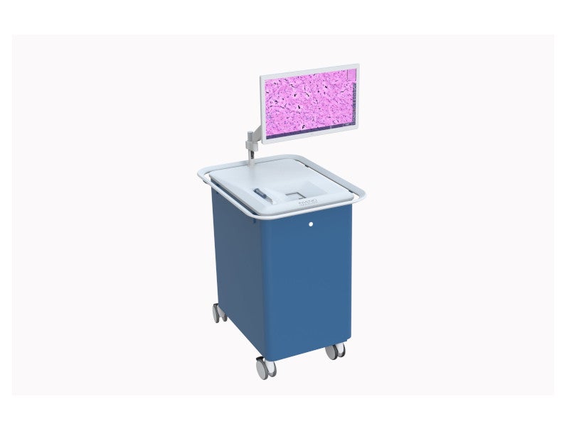

The NIO laser imaging system is manufactured by Invenio Imaging, a medical device company based in California, US. It produces anatomy images for use during surgical procedures.
The device received CE mark in September 2021, meaning that it complies with high health, safety and environmental protection requirements.
Recommended Buyers Guides
The approval allowed Invenio Imaging to commercialise the imaging system for intraoperative histology in the European market.
Invenio Imaging partnered with Ambra Health in July 2020 to integrate the NIO laser imaging system with the latter’s Cloud PACS image management technology and viewing capabilities.
The integration enables pathologists to instantly access Stimulated Raman Histology (SRH) images exported from Invenio’s imaging device. The Cloud technology allows pathologists to access images from anywhere with minimal delay.
The NIO laser imaging system has been used in more than 2,500 treatments in major cancer centres across the US and Europe.
NIO laser imaging system features
The NIO laser imaging system can capture real-time microscopic images of fresh tissue specimens intraoperatively in less than three minutes, which helps to provide quicker feedback to surgeons.
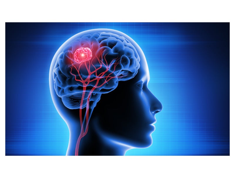
It enables the intraoperative diagnosis of brain tumours. The system, in combination with 5-ALA fluorescence and other essential imaging techniques, can improve the detection of brain tumours during surgery.
A customised NIO Slide can be used for squash preparation and to retrieve samples for subsequent downstream analysis after imaging.
The NIO laser imaging system streamlines intraoperative histology and reduces operating room (OR) downtime.
It allows surgeons to instantly access gold-standard tissue histology at the time of surgery, allowing them to make more informed surgical decisions. It also streamlines intraoperative pathology consultations.
The system uses a high numerical aperture objective with a 25x magnification and 0.5mm scan width. Furthermore, it features a versatile interface for image sharing and archiving.
Imaging procedure
The NIO system uses SRH and Stimulated Raman Scattering Microscopy to provide microscopic images.
It can provide images of unprocessed tissue specimens without physical sectioning or dye staining, enabling histologic evaluation without the need for a laboratory.
SRH uses optical sectioning to create three-dimensional images of thick specimens. It leverages laser spectroscopy to examine the sample’s chemical makeup and enables optical imaging of fresh tissue specimens without the need for elaborate tissue preparation.
SRH can transform digital pathology and allow pathologists to see cellular characteristics and architecture in real-time.
NIO Glioma Reveal image analysis module
Invenio Imaging received CE mark for the NIO Glioma Reveal image analysis module in May 2022.
Using artificial intelligence (AI), the NIO Glioma Reveal image analysis module generates microscopic anatomy images for use during surgical procedures, which can help in the detection of cancer during surgery.
It is used by neurosurgeons to spot cancer infiltration in patients with diffuse glioma undergoing primary treatment.
Diffuse gliomas
Diffuse gliomas are one of the most common types of brain tumours and generally affect the cerebral hemispheres of adults. They are classified under different grades based on the histologic degree of malignancy.
The prognosis of diffuse gliomas depends on the histologic grade. Diffuse midline gliomas, a subtype, are primary central nervous system tumours that begin in the brain or spinal cord.
They most commonly originate in the pons, located in the brainstem, thalamus, cerebellum and spinal cord.