
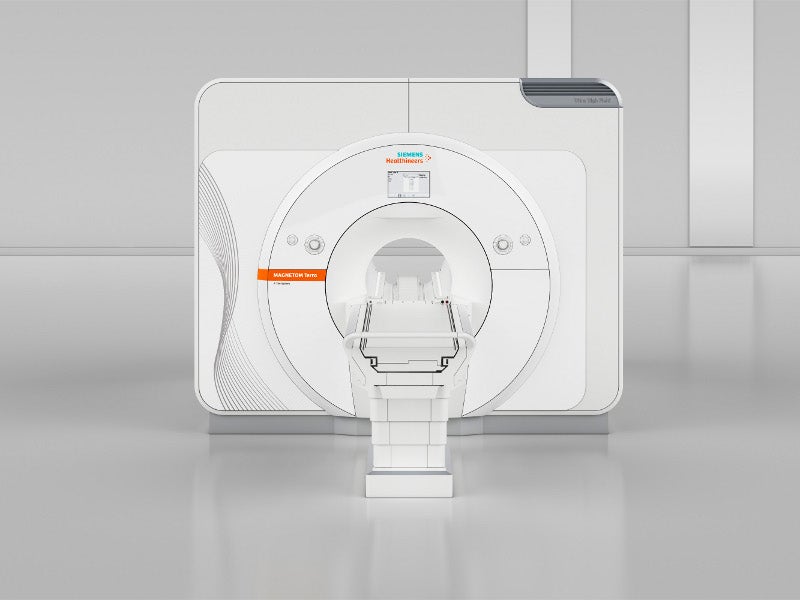
The Magnetom Terra 7 Tesla (7T) magnetic resonance imaging (MRI) scanner is one of the first 7T MRI scanners cleared for clinical imaging in the US and Europe.
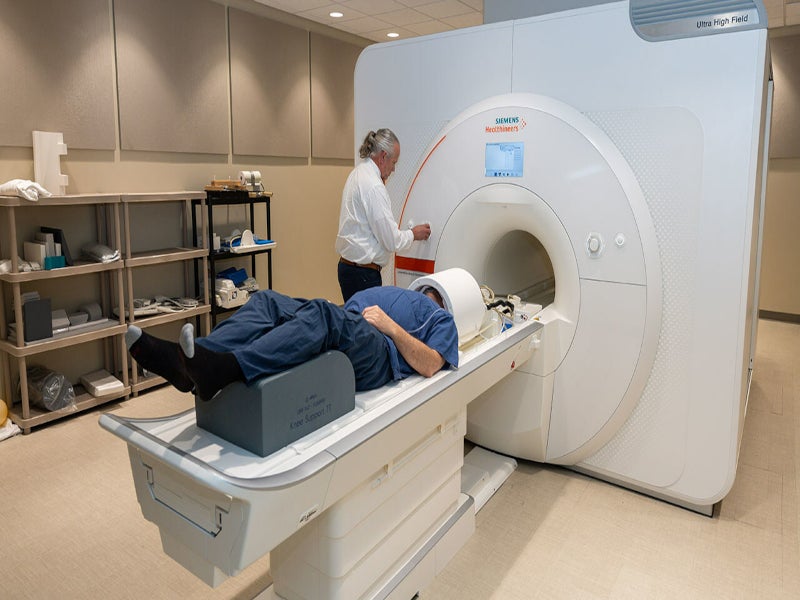
Developed by Siemens Healthineers, the device is designed to produce cross-sectional images of the brains and knees of patients weighing 66lbs (30kg) or more.
Recommended Buyers Guides
The Mayo Clinic in Rochester, Minnesota, US, was one of the first medical institutes in North America to use the Magnetom Terra for clinical practice.
The machine is installed at Brigham and Women’s Hospital (BWH) in Boston, Massachusetts and the Keck School of Medicine’s Mark and Mary Stevens Neuroimaging and Informatics Institute.
In September 2019, the University of Liege installed the first operational 7T MRI scanner at the GIGA-Research Centre of the Cyclotron-In Vivo Imaging of ULiege in Belgium. The state-of-the-art technology enables researchers to understand the physiology and brain pathology of neurological diseases such as Alzheimer’s disease, epilepsy, Parkinson’s disease and multiple sclerosis more precisely.
The machine arrived in Trondheim, Norway, in September 2019. It is part of the NORBRAIN initiative and is managed by the Norwegian 7T MR Center.
Magnetom Terra X is the next-generation FDA-cleared 7T MRI. The system was installed in the Auburn University Samuel Ginn College of Engineering’s Neuroimaging Center in March 2024 for use on clinical patients.
Regulatory approvals for Magnetom Terra 7T scanner
In August 2017, the scanner obtained Conformité Européene (CE) and 510(k) certifications for clinical use in Europe.
The US Food and Drug Administration (FDA) approved the device for clinical imaging in October 2017.
Magnetom Terra 7T scanner technical details
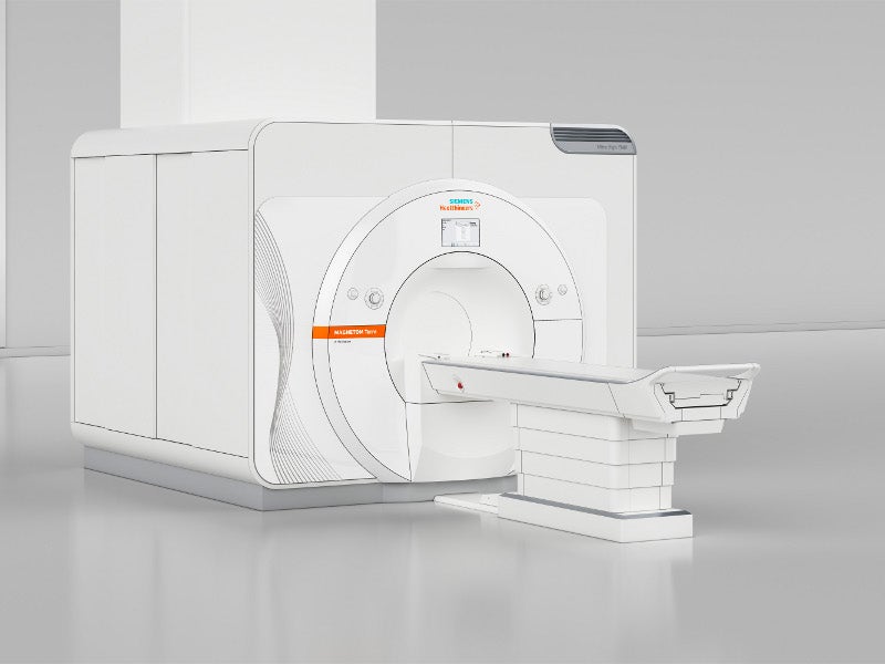
The Magnetom Terra 7T MRI scanner measures 2.97m in length and occupies a 65m² (699.6ft²) area. It boasts an ultra-high magnetic field strength of 7T, 140,000 times more powerful than the Earth’s magnetic field.
The advanced system is designed to provide exceptional image resolution due to its zero helium evaporation technology, which employs helium to cool the magnet to -270°C, enabling superconductivity and the generation of the 7T magnetic field.
Weighing less than 25t, the scanner’s actively shielded, highly homogeneous superconducting magnet is 50% lighter than comparable 7T magnets. With a bore size of 0.6m and a length of 2.7m, it is uniquely transportable in a cold state by aircraft.
The Magnetom Terra 7T’s open system architecture incorporates two coils for precise imaging of the head and knee, and its 80/200 gradient system supports advanced techniques such as diffusion MRI and functional MRI (fMRI).
The system’s eight-channel parallel transmission (pTX) technology enables the capture of detailed images from challenging body regions, and it can be expanded to a 16-channel pTX for even greater flexibility.
With up to 64 receive channels, the scanner delivers an ultrafine 0.2mm in-plane anatomical resolution, enhancing the visibility of small lesions and potentially aiding in the diagnosis of previously undetected conditions.
The Magnetom Terra 7T offers a double signal-to-noise ratio for increased precision in clinical applications and features Dual Mode functionality, which allows for a secure switch between research and clinical operations in under seven minutes.
Additionally, its software platform facilitates the easy sharing of study protocols with other magnetic resonance systems in clinical routine, enhancing its utility in both research and clinical settings.
Magnetom Terra 7T scanner applications
The Magnetom Terra 7T scanner is a cutting-edge device that supports basic clinical research, particularly in musculoskeletal and neurological fields. It enhances the visibility of minute pathologies, facilitating detailed anatomical imaging of cells in musculoskeletal conditions and enabling the examination of sub-cortical brain activity through functional MRI techniques.
The high-resolution imaging capability serves as an MRI microscope, allowing for the exploration of metabolic changes by assessing the anatomy, function and metabolism of body tissue. The results obtained from the scanner are instrumental in guiding treatment decisions and evaluating treatment efficacy. Additionally, the Magnetom Terra has a flexible design, making it adaptable for a range of future clinical applications.
Magnetom Terra 7T scanner benefits
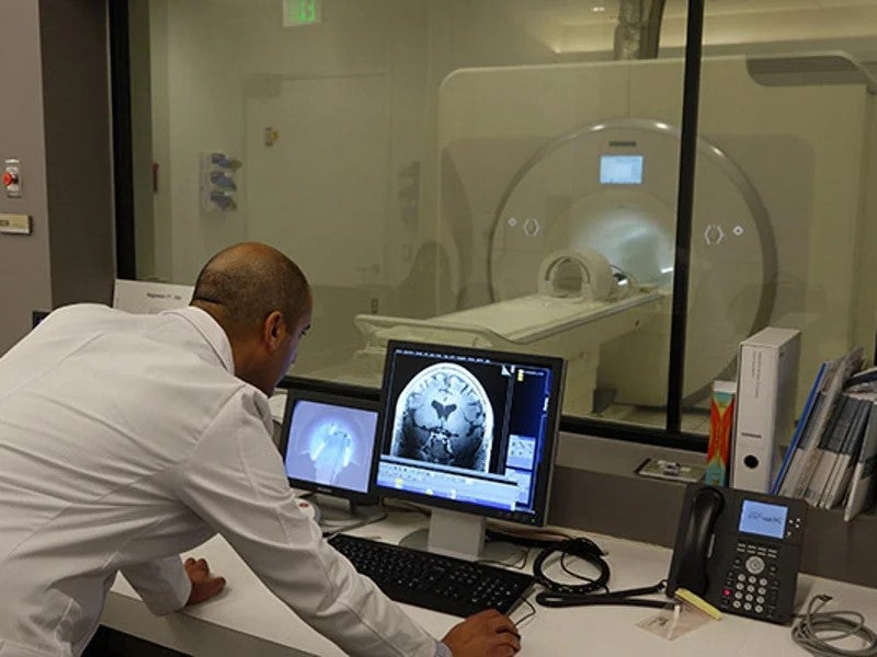
The Magnetom Terra 7T MRI Scanner is a state-of-the-art device capable of producing high-resolution images as small as 0.14cm³, enhancing the visibility of lesions for metabolic brain mapping.
Its sub-millimetre BOLD fMRI precision enables the visualisation of sub-cortical activations, with fMRI technology measuring changes in blood flow and oxygenation to provide insights into brain activity.
The heightened sensitivity of the 7T system allows researchers to detect subtle changes in brain function, facilitating more accurate mapping of brain regions associated with various cognitive processes. The feature is particularly significant for studying conditions, such as Alzheimer’s disease, stroke and psychiatric disorders.
The scanner’s versatility extends to both research and 510(k)-cleared clinical modes, supporting clinical routines and pioneering translational research. It is instrumental in rapidly identifying biomarkers for disease symptoms, elucidating metabolic processes, understanding sleep mechanisms in the brain and visualising neurodegenerative diseases.
Magnetom Terra X details
Magnetom Terra X utilises groundbreaking Ultra IQ technology. The updated version features new hardware and XA60A software.
Ultra IQ technology offers unprecedented image clarity due to its dynamic parallel transmit architecture, which enables confident assessment of the most subtle pathological details.
Magnetom Terra X elevates clinical routines to new heights by combining 7T field strength with the enhanced performance of AI-powered image reconstruction Deep Resolve. The integrated Open Recon3 platform enables the implementation of custom reconstructions directly on the scanner to ensure a smooth transition between research and clinical workflows.
The Magnetom Terra.X allows for a complete brain and knee exam with unparalleled resolution in under 15 minutes of scan time.