
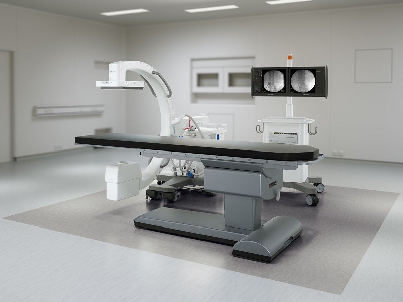
Philips Image Guided Therapy Mobile C-arm System 1000 – Zenition 10 is an innovative surgical imaging system developed by Royal Philips, a health technology company based in the Netherlands.
The system incorporates enhanced features to expand the imaging capabilities of surgeons and improve operating room (OR) performance in a wide range of routine surgical care and minimally invasive procedures, including orthopaedic, trauma, spine, pain management, peripheral vascular, abdominal, urology and general surgical procedures.
Recommended Buyers Guides
The Zenition mobile C-arm imaging platform was initially launched in the US, Germany, Austria and Switzerland in February 2019 to provide live image guidance during a wide range of surgeries for improved clinical accuracy and efficiency.
Launched in May 2023, The Philips Zenition 10 is a new addition to the existing Zenition mobile C-arm series, which includes the Zenition 50 and Zenition 70 systems. Philips’ Zenition C-arms are CE marked and have received the US Food and Drug Administration (FDA) clearance for human use.
Zenition 10 image-guided mobile therapy mobile C-arm design and details
The Philips Zenition 10 is a state-of-the-art surgical imaging system that combines a monoblock 40KHz high-frequency generator with a maximum output of 2.1KW and a fixed anode X-ray tube for advanced X-ray generation. It is equipped with a state-of-the-art flat detector with Trixell amorphous silicon technology to deliver high-quality imaging, high uptime, and efficient workflow.
The detector area measures 20cm x 20cm, with FD11: 1024 x 1024 pixels matrix size and pixel pitch set at 200µm, providing a wide coverage area for capturing distortion-free images with a smaller pixel pitch for higher spatial resolution.
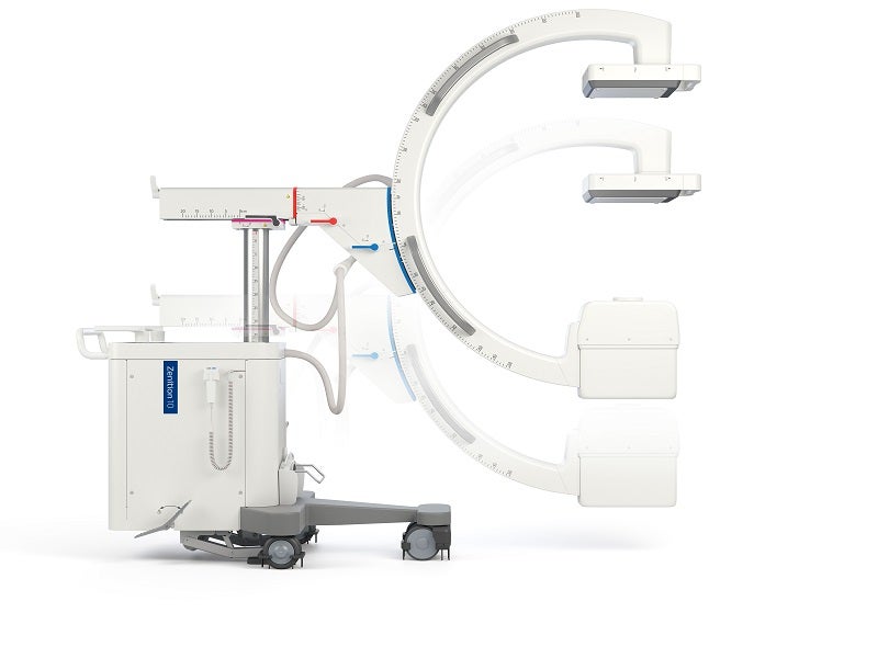
The company’s flat detector technology with fixed and mobile C-arm systems offers a compact design to perform the daily mix of surgical procedures. The C-arm stand is 2,138mm long, 850mm wide and 1,640mm high, and weighs up to 330kg. It can rotate more than 180° and pan more than 12.5°. The exceptional C-arm geometry with a depth of 730mm and angulation movement of 150°, together with the compact flat detector, enables quick and convenient positioning around patients. The source-to-image distance (SID) of the C-arm stand is 1,000mm.
As part of the Zenition series of mobile C-arm systems, The Zenition 10 offers features, including C-arm manoeuvrability, one-click application-specific protocols and more customisation with user profiles. It also offers a dedicated low-dose paediatric mode for young patients.
The Zenition 10’s mobile C-arms can be easily connected to the hospital infrastructure through advanced connectivity and interoperability tools, including high-speed wireless data transfer and full digital imaging and communications in medicine (DICOM) 3.0 capability.
Features for improved user experience
The Zenition 10 offers ease of use and operation with improved accessibility to anatomy. The space-saving tank design offers more space for easy manoeuvrability of the C-arm between the tank and the floor or table base.
The system can perform more flexible movements with the fully counterbalanced C-arm and the lightweight mobile viewing station measuring 830mm deep, 830mm wide and 1830mm high, with monitor height movement up to 150mm.
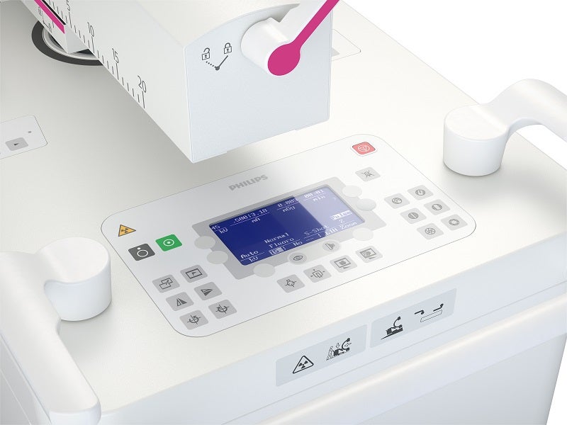
The navigational aids based on the principles of Unify workflow made imaging easier with intuitive control and handling, which reduces miscommunication while improving teamwork. Some features of the Unify workflow include ClearGuide and Color Coding.
Its uniform design and user-friendly controls make users feel at ease while using the system, which helps to simplify use and streamline fleet management.
The system helps avoid operational delays, making it suitable for emergency scenarios and in multiple areas of a hospital. The large storage capacity of 140,000 images also avoids operational delays from a lack of image spacing.
The Zenition 10’s digital subtraction angiography capability further improves surgical procedures by enabling clear imaging of vasculatures. It has several real-time processing and post-processing functions for improved imaging.
Additional features of Zenition 10 mobile C-arm system
Proactive logging and Philips’ Remote Expert Connect features reduce surgical interruptions with planned downtime. The same features also help in planning the operational costs more effectively.
Philips’ Hospital Operational Services (HOS) customised service agreements help Zenition 10 to maintain productivity high with minimal effort.
Intuitive operating controls, on-screen help, digital user guides, and clinical education solutions promote a fast learning curve and reduce the operating staff’s training time.
The Zenition 10 offers an uninterrupted power supply for free movement from one operating room to another without rebooting the entire system. The standard Windows® platform offers enhanced cybersecurity to the system.
Other tools and technologies
The Zenition 10 uses advanced image processing algorithms combined with Philips’ DoseWise radiation dose management features to offer high-resolution images at efficient dose levels.
The MetalSmart option minimises the impact of metal objects within the field of view to improve the image quality for patients undergoing orthopaedic procedures or those with metal implants.
BodySmart software consistently delivers high image contrast by automatically adjusting the measuring field accurately to the area of interest, promoting dose efficiency and first-time-right imaging.
The Multi-Modality Viewer enables the upload of computed tomography (CT), magnetic resonance imaging (MRI), and other DICOM images to compare with live fluoroscopy images side by side for more precise clinical decisions. The outlining tool helps to mark a bifurcation, side branches, or any other anatomy on live fluoroscopy images for guidance during surgical procedures.