
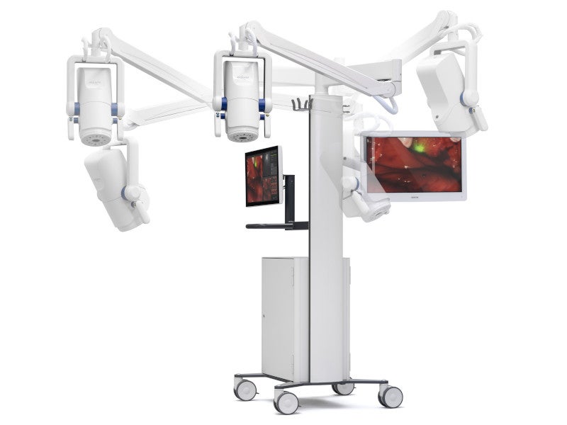
The Explorer Air II is an intra-operative imaging system developed by SurgVision, a medical device company based in the Netherlands. It enables surgeons to visualise fluorescence in the near-infrared (NIR) range inside the body of a patient, allowing real-time imaging during surgery.
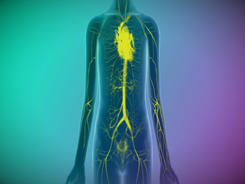
Based on SurgVision’s next-generation technology platform, the system is used for the visual assessment of blood flow and tissue perfusion before, during and after vascular, organ transplant, plastic, gastrointestinal, and micro and reconstructive surgeries in adult and paediatric patients aged one month and older.
Recommended Buyers Guides
It is designed to fulfil the requirements of oncological intraoperative fluorescence imaging by offering exceptional sensitivity and imaging accuracy.
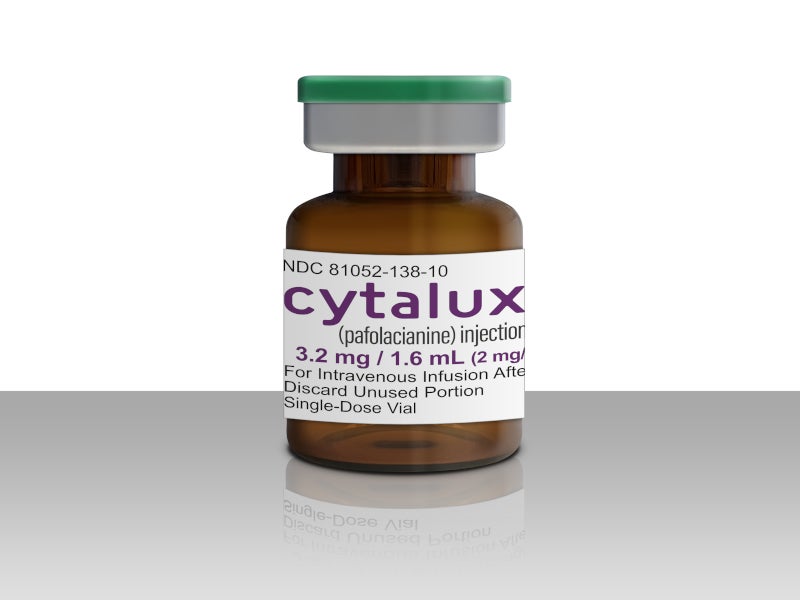
The development of the Explorer Air II system was started in 2019. The system received 510(k) clearance from the US Food and Drug Administration (FDA) for indocyanine green (ICG)-based perfusion imaging in 2022. The indication of use was expanded with pafolacianine for intraoperative fluorescence imaging of tissues in March 2023.
The system was also CE-marked for perfusion imaging in the EU.
SurgVision’s Explorer Air II design and features
The Explorer Air II imaging system consists of two cameras, one for fluorescence and the other for coloured images, suspended by an articulated arm attached to a trolley. A touch screen and secondary monitor are also mounted on the trolley.
The tissue of interest in the patient’s body gets illuminated upon intravenous administration of fluorescent dye, such as ICG. The device uses a combination of NIR light and advanced imaging techniques to visualise fluorescence excited by infrared light ranging between 740nm and 760nm and emitted in the band centred around 800nm.
The infrared light absorbed by the tissues and blood vessels in the surgical field allows the camera system to capture images of the tissues and vessels that are not visible to the naked eye.
The images captured by the camera system are then processed and displayed in real time using a software platform. It allows the surgeons to locate the blood vessels, tissue perfusion, and tissue oxygenation in real time during surgery, providing them with valuable information that can help guide their decisions and improve surgical outcomes.
The composite image is displayed along with the fluorescent and colour images, to tag and compare them, play the recorded videos, and export selected files.
It is recommended to use the Explorer Air II with Explorer Air Sterile Drape under sterile conditions. The sterile drape is a single-use, custom surgical drape attached to the camera head to maintain the sterility of the operating field throughout the procedure.
SurgVision’s Explorer Air II benefits
SurgVision’s Explorer Air II enables reliable detection of lesions less than 3nm in a lower tracer dose. Its high fluorescence frame rate allows seamless real-time imaging. The device allows the display of three images, colour, fluorescence, and overlay, simultaneously.
Its dynamic range allows to image high and low levels of fluorescence within one image session. It helps in imaging different intensities of fluorescence over time.
The LED-based excitation prevents the use of a high-power laser. The system offers consistent fluorescence during procedures and homogenous illumination of the field of view.