
A mass spectrometry technique developed at University College London (UCL) is enabling cancer researchers to analyse the individual behaviour of millions of different cells inside lab-grown bowel tumours.
The research, which has been published in Nature Methods, provides new insight into how mutated cancer cells mimic the growth signals expressed by healthy cells. This could help scientists understand how tumours are able to evade the immune system and become resistant to treatments.
Go deeper with GlobalData

Discover B2B Marketing That Performs
Combine business intelligence and editorial excellence to reach engaged professionals across 36 leading media platforms.
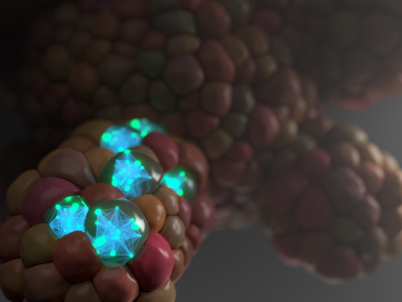
The 3D lab-grown tumours, known as organoids, are grown by embedding cancer stem cells in collagen. They contain many different cells and more accurately represent a patient’s cancer compared to more traditional research, which looks at a collection of identical cells grown in 2D.
However, most methods to study organoids involve grinding up the cells and analysing the resulting mixture. This limits researchers’ ability to assess how individual cells behave or how tumours collaborate with healthy and immune cells known to influence cancer outcomes.
Researchers at UCL have now developed a new mass spectrometry technique to measure communication signals in millions of single cells in bowel organoids.
At a subcellular level, heavy metal-tagged antibodies are attached to individual proteins, known as post-translational modification (PTM). The different weight of each heavy metal allows the mass spectrometer to differentiate cell proteins and analyse their behaviour and signalling activity, both within and between different cell types.
More than 40 types of PTM were tagged in the process, enabling the researchers to build detailed circuits that describe how cancer cells are working. They were able to detect 28 key signalling molecules across six different cell types in over one million cells.
The study’s findings showed that cancer cells themselves, as well as immune cells and connective tissue, rewire the normal signalling networks of bowel tissue, allowing tumours to grow freely.
UCL Cancer Institute principal research fellow and the study’s corresponding author Dr Chris Tape said: “Organoids are already revolutionising cancer research by allowing us to test whether experimental new drugs are effective on lifelike models of tumours. But crucially, this new technique helps scientists to understand why a treatment works or not, by revealing in unprecedented detail how cells are talking to each other.”
Moving forward, UCL scientists plan to use this technology to study how tumours from individual patients can uniquely communicate with healthy cells and the immune system.
It is hoped the research will open the door for the development of more effective drugs, by revealing why tumours respond to treatments in the way they do. It could also help doctors to select the best course of treatment for individual patients, as they could grow an organoid of their tumour and test different treatments on it before prescribing them.
