
Researchers at the National Eye Institute (NEI), part of the US National Institutes of Health (NIH), have used a new imaging technique to reveal important details regarding choroideremia, a rare eye disease.
They combined traditional eye imaging approaches with adaptive optics technology, which improves the resolution of the image.
Go deeper with GlobalData

Discover B2B Marketing That Performs
Combine business intelligence and editorial excellence to reach engaged professionals across 36 leading media platforms.
The research has revealed how the cells in different tissue layers in the eye are affected in choroideremia patients.
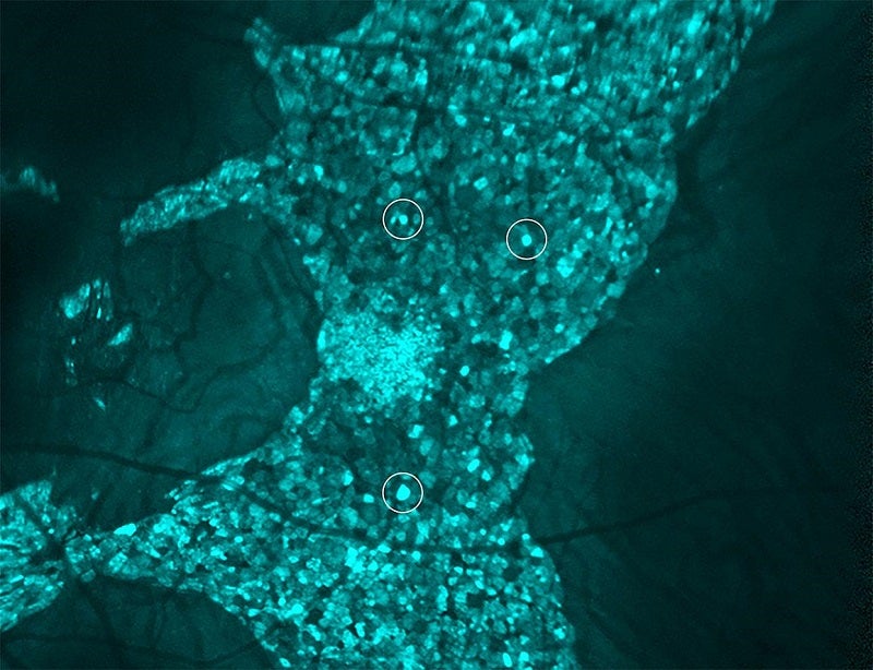
NEI Clinical and Translational Imaging Unit head Johnny Tam combined adaptive optics and indocyanine green dye to see live cells, including retinal pigment epithelial (RPE), choroidal blood vessels and light-sensing photoreceptors, found in the retina.
Through this approach, Tam’s team was able to see the extent to which choroideremia damages the tissues.
This information may help in designing effective treatments for choroideremia, as well as other diseases.
Tam said: “One major finding of our study was that the RPE cells are dramatically enlarged in males and females with choroideremia.
“We were surprised to see many cells enlarged by as much as five-fold.”
In the study, published in Communications Biology, female participants had both enlarged and healthier-looking RPE cells.
According to Tam, this could explain why women with choroideremia have fewer symptoms.
In both male and female participants of the study, photoreceptor and blood vessel layers were less affected, which suggested that disruption of RPE plays a key role in choroideremia.
Tam added: “It’s not obvious at first, but using an existing tool in the clinic, we can monitor and track the cellular status of the RPE layer.
“This could prove valuable in identifying which patients would benefit the most from therapeutic interventions.”
The NEI Intramural Research Program provided funding for the study.
